In order for tumor cells to multiple and spread, they must develop their own blood supply through a process called angiogenesis. Angiogenesis inhibitor chemotherapy drugs work to stop or slow down this process, thereby controlling tumor growth. Metronomic chemotherapy is one example of angiogenesis inhibition treatment, which is becoming a popular treatment option for pets with cancer.
The definition of metronomic chemotherapy is variable, but usually refers to the continuous administration of low doses of oral chemotherapy drugs designed to target the endothelial cells lining the blood vessels supplying tumor cells.
When traditional cytotoxic chemotherapy is administered at maximally tolerated doses (MTD – see previous blog article entitled “Is the remedy for cancer worth the cure?”), death of the endothelial cells lining the blood vessels of tumor cells occurs first, followed by the death of the tumor cells. When we administer chemotherapy in this manner, we typically need to give…
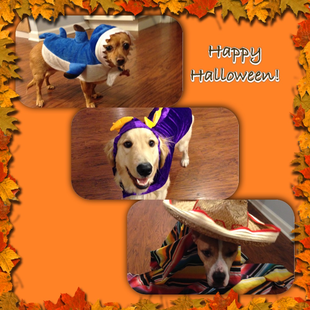
As Halloween and the upcoming holidays are rapidly approaching, we are often wrapped up in family gathering, parties, and other activities and forget about the well being of our beloved pets. I tried compiling a short list to help keep our pets healthy and out of trouble this holiday season. This should ensure that everyone has a happy holiday season and may save you and your pet from needing emergency trips to veterinarian or emergency clinic.
1. Keep candy away from pets, in particular chocolate and candies made with xylitol and other sweeteners. While these taste good, they can have very harmful effects on your pet, ranging from liver failure, seizures, and as severe as death. If you suspect your pet has consumed any of these, seek veterinary care immediately.
2. Keep a close on the whereabouts of your pets. With all the excitement and increased visitors during this time, make sure your pets are accounted for and haven’t run off. Missing pets and subsequent trauma, such as being hit by a vehicle is an all to common occurrence during this time. Make sure your pets are in a safe place when company is over.
3. Keep pets out of the garbage and from grabbing food off the table. Bones and fatty meats can cause illness in our pets, especially dogs. Bones can cause a lot of irritation and in some cases puncture the gastrointestinal tracts. Fatty foods are not good for our pets and can cause pancreatitis and other gastrointestinal issues. Pancreatitis can range in severity and needs to be treated by your veterinarian.
4. Keep cords and electrical wiring away from your pets. Both cats and dogs can find wires enticing. Electrocution injury can be very severe and cause death in some cases. If you believe your pet to have be electrocuted, have them evaluated by your veterinarian immediately.
5. Keep easily ingestible objects away from your pets. Objects that can be easily swallowed can cause gastrointestinal irritation and obstruction. Some objects that can become obstructive are clothes, small toys, tinsel, etc. Gastrointestinal obstructions demand immediate veterinary care. There are times when the object can pass, but most of the time your pet will need surgery to relieve the obstruction, Surgery can range from a single incision in the stomach to removal of a segment of intestine. In extreme cases this condition can be fatal.
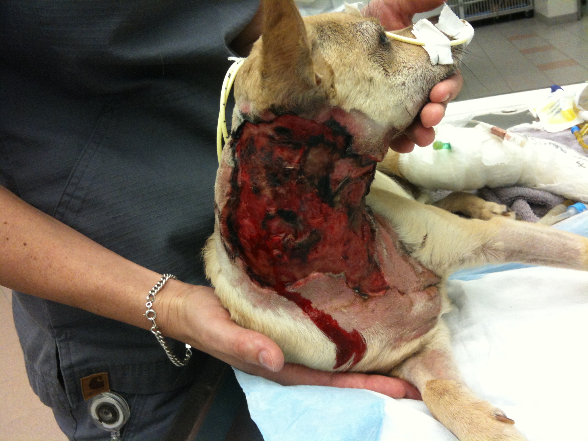
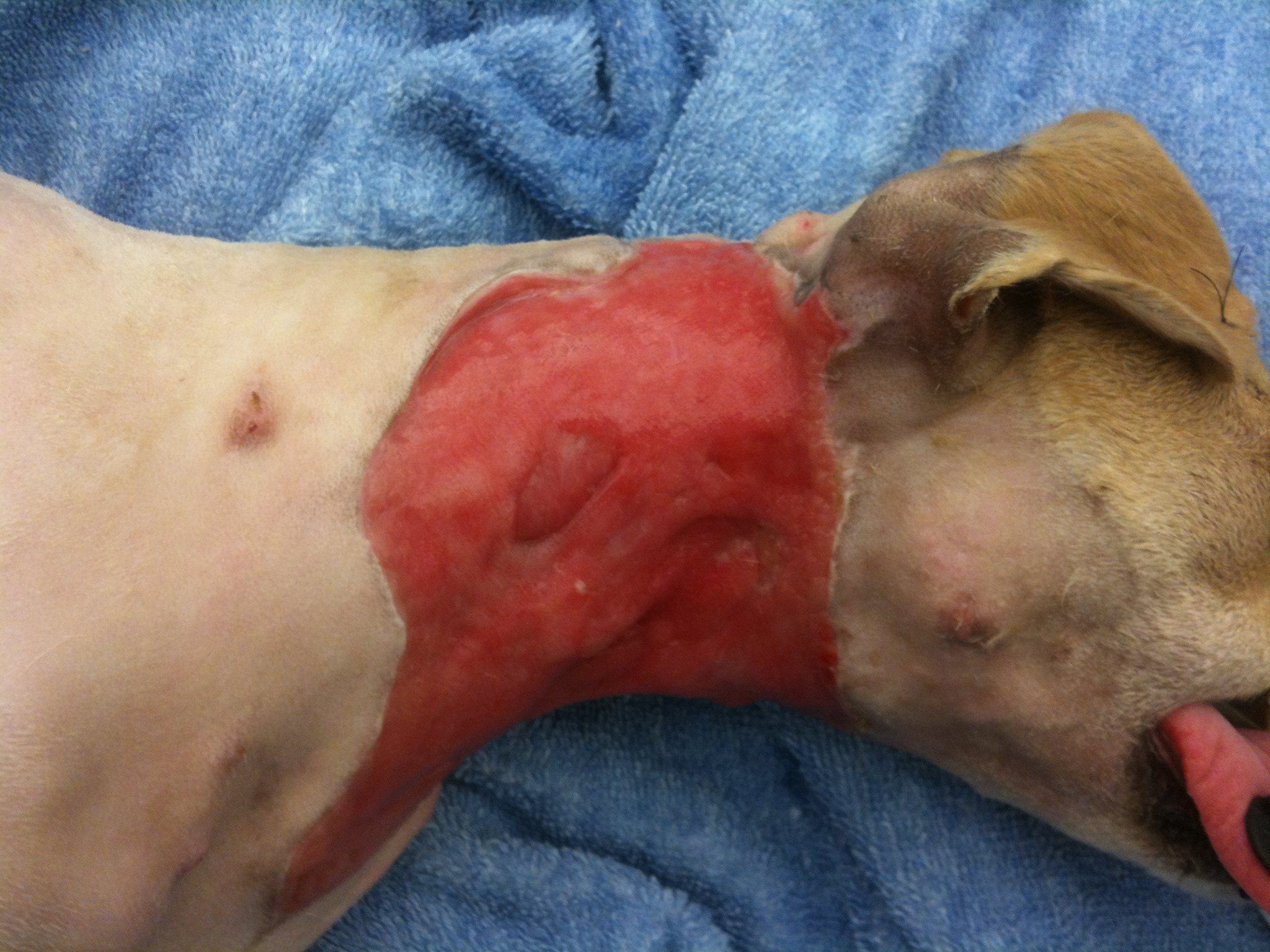
October’s case of the month is an interesting one. Lucky is a young chihuahua mix that came to us with SEVERE injuries. He was attacked by a neighborhood dog about 7 days before presenting to us and was in very bad shape. His initial wounds were managed, but unfortunately infection still set in and the majority of his cervical (neck) skin started to die and needed to be removed. Not only was the infection causing a problem locally, but we had signs of it being spread systemically (through his blood stream). He needed both surgical care and care by a criticalist(board certified in Emergency and Critical Care) in order for him to have a fighting chance. Below is a picture of what he looked like when he was admitted into the hospital.
This was Lucky as he was admitted to the hospital.
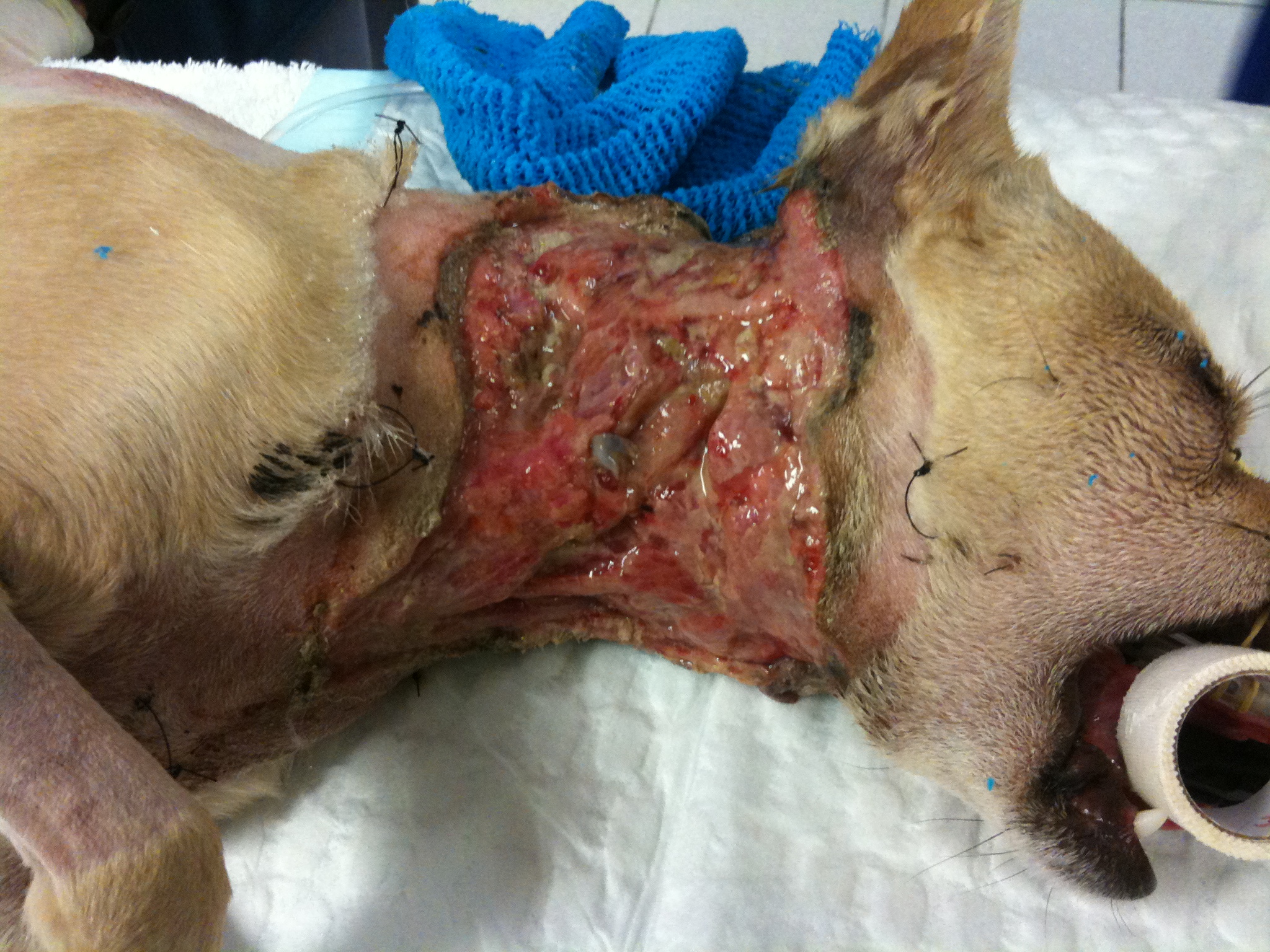
You can make out the extensive injuries on the photo above. He was fortunate to be alive! Whenever we get a case like this (unfortunately it happens more frequently than we like), we am always realistic with the owners, because there is a chance that their pet will not survive. Also, these cases are not quick cases, Lucky was hospitalized for 2-3 weeks and wasn’t fully healed for about 6 weeks. Our first objective is to get the systemic infection under control and get him strong enough to be able to handle surgery. While the criticalist was working on the systemic infection, we were concentrating on the neck wound.
The first phase wounds go through is the debridement phase, which is where the body gets rid of necrotic (dead) tissue and the size of the wound is established. The next phase to follow is the granulation phase. The granulation phase is very important in a large wound like this, this is when the body begins to infiltrate the wound with healthy tissue and more importantly capillary vessels, which bring blood flow. For this wound, I choose to use a wound dressing called BurnStat (Ubuntu) which is a dressing that can be used through multiple phases. It is an organic clay substrate that does an excellent job of removing toxins and necrotic tissue while promoting granulation tissue formation.
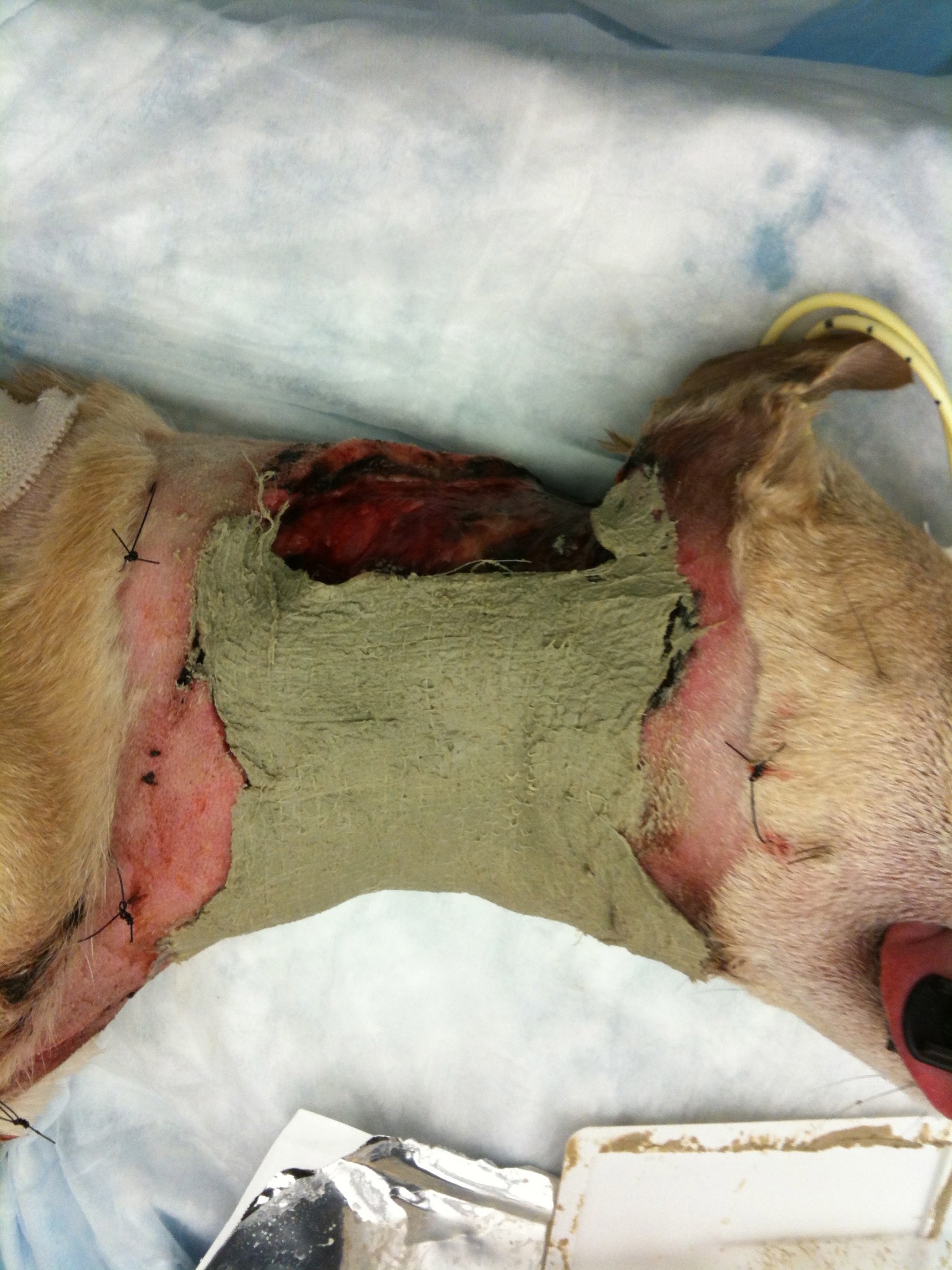
Debridement phase
Applying BurnStat as the primary wound dressing
Following this type of bandaging, the diseased tissue begins to be replaced with more healthy, red granulation tissue. The final product, before being able to close the wound, needs to be completely covered with granulation tissue in order to increase the chance of the new skin being accepted.
Complete coverage by granulation tissue.
You can see how the surface is covered with healthy looking tissue and no presence of dying tissue visible. By this time Lucky amazingly over came his battle with the widespread infection and overall was doing very well. He was making a remarkable recovery.
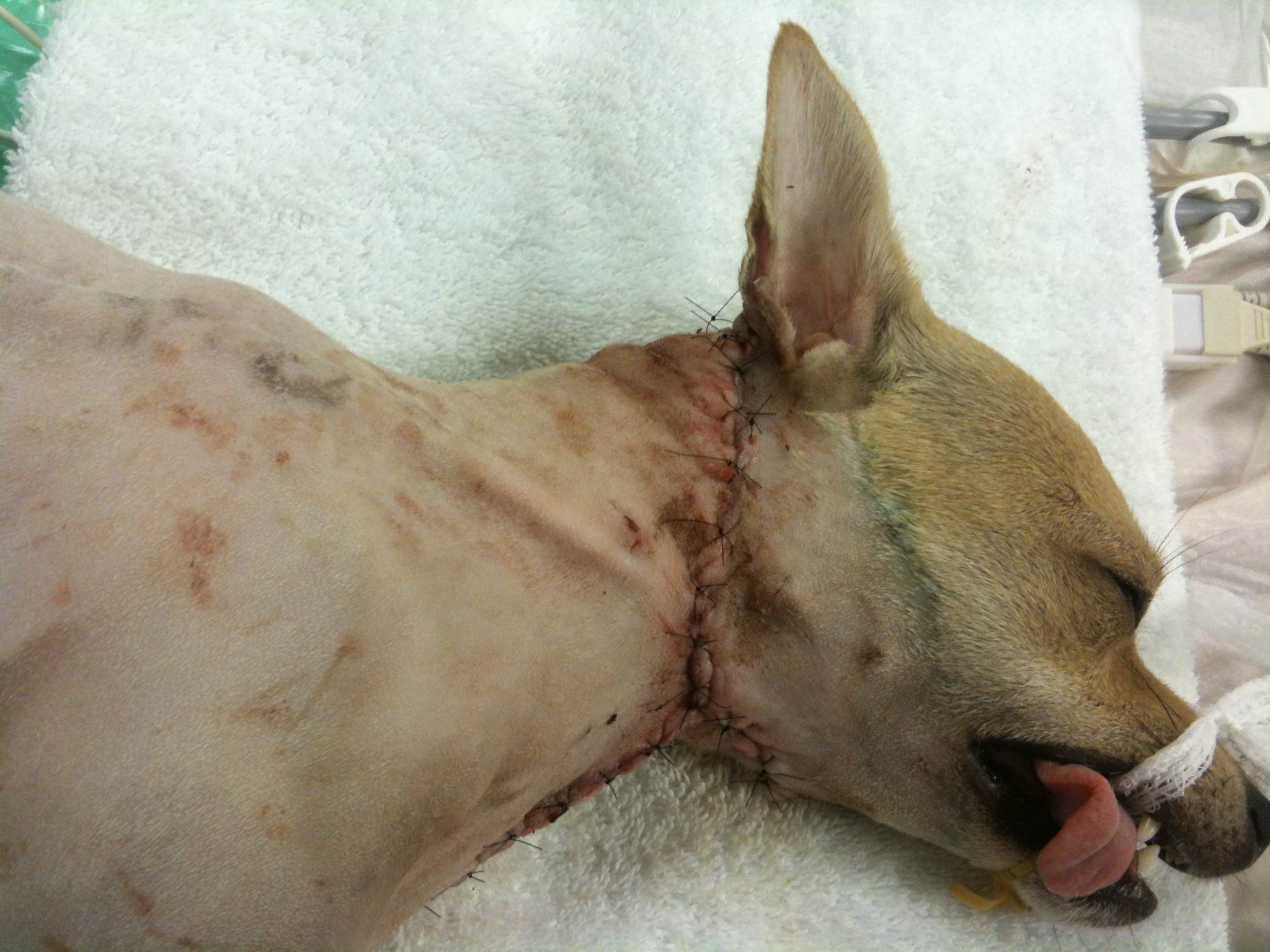
Our next dilemma was “how do we cover the exposed tissue”? In cases like this, we have a few options, which is beyond the scope of this post. I choose to use an advancement flap (skin freed up from a nearby location that is moved over the wound), which made the most sense due to the elasticity of the skin in this area. Below is his wound following surgery.
Advancement flap
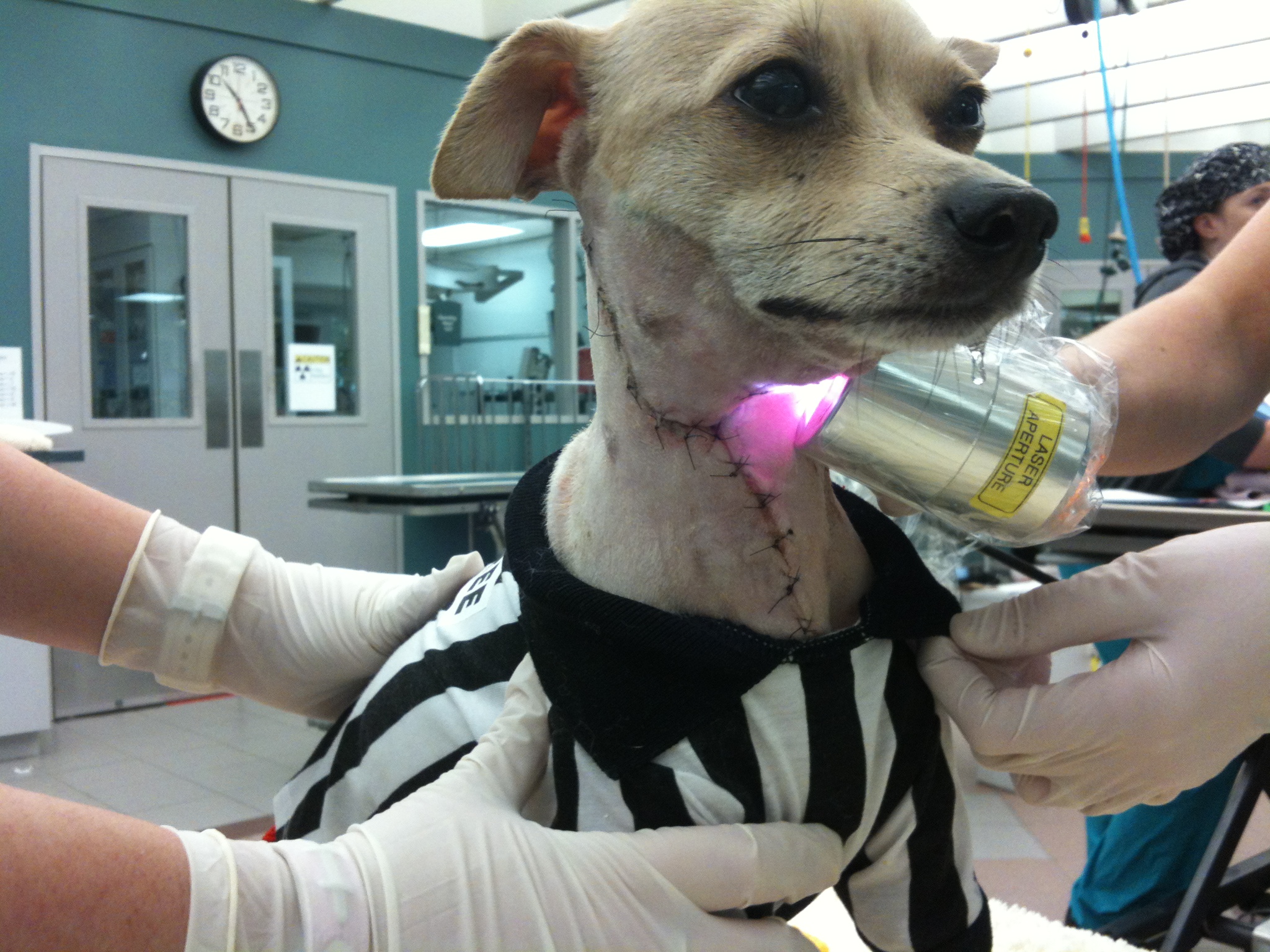
Following surgery we also utilized laser therapy to help promote uptake of the skin flap. This is Lucky in his referee uniform (it is close to Halloween) receiving his laser therapy.
Post-advancement flap therapy
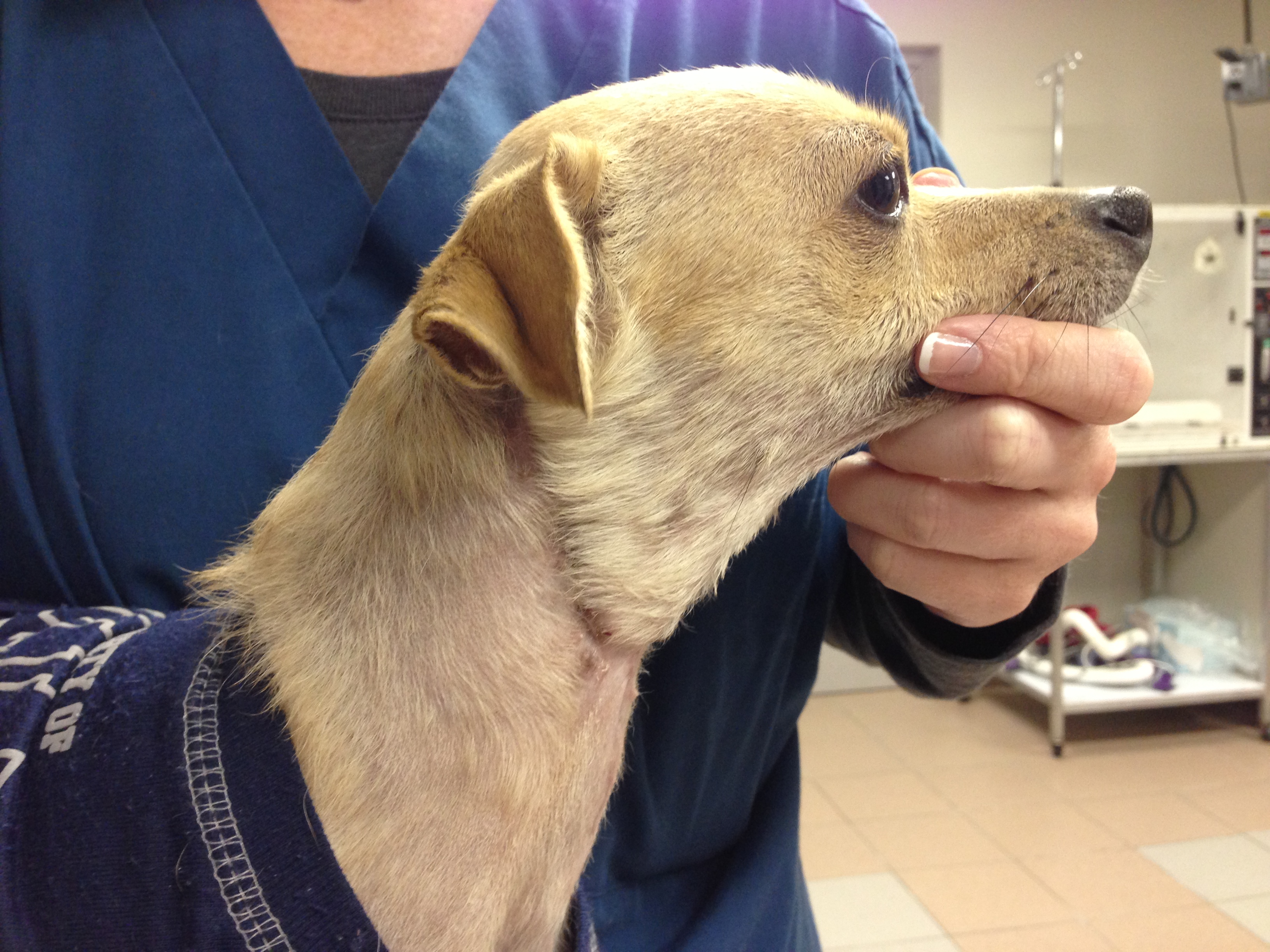
Here is the finished product for Lucky!!! He overcame a lot of obstacles along the way!!
Very good article to help explain common terms used in medicine. Dealing with cancer in a pet is troubling enough (having gone through it with my own dog), not to mention getting bogged down with new words and terms. I hope everyone will find this beneficial.
Chemotherapy and radiation therapy are confusing topics. When complicated terminology is combined with the anxiety associated with a diagnosis of cancer, it’s easy to understand how things become blurry. Further complicating things are those veterinarians who cross specialties. How can an owner keep be expected to keep it all straight?
Chemotherapy is defined as the use of chemical substances to treat disease. Conventionally, we think of chemotherapy in relation to treating cancer. Chemotherapy can be administered intravenously (through a vein), topically (on the skin), subcutaneously (under the skin), intramuscularly (into a muscle), orally, intratumorally (injected directly into a tumor), or intracavitary (given directly into a body cavity.)
Adjuvant chemotherapy is prescribed after a tumor is removed and we are hoping to treat any microscopic residual cancer cells that may have spread from the tumor prior to surgery. An example of adjuvant chemotherapy is treating a dog with osteosarcoma with a…
Sometimes it can be very hard to determine which leg your pet (or patient) is limping on, let alone which joint is causing the problem. I want to take a little time to discuss a problem that we see from time to time that typically affects the juvenile (6-18 month), medium and large breed dog and is typically thought of as a congenital/hereditary issue. The most note worthy joints affected are the shoulder (proximal humerus), the elbow (distal humerus), stifle (distal femur), and hock (talus).
The underlying etiology is similar in all the joints, however this article will focus on the shoulder with subsequent articles dealing with the other joints. I think this approach is reasonable because the treatment may be different for other joints,as well as, the prognosis can vary. Again, this disease affects primarily young dogs; in the older patients we usually see the consequence of this issue, resulting in osteoarthritis of the joint.
Osteochondrosis (OC) precedes osteochondritis dissecans (OCD) and is characterized by a problem between the metaphyseal growth plates of the affected bone and the cartilage. In essence, the cartilage surface does not adhere to the underlying subchondral bone surface. When a cleft or break develops in this “soft” cartilage, this fulfills the term OCD. Once the area progresses to an OCD lesion (a break in the cartilage develops), then the patient becomes clinically lame and will exhibit a degree of lameness/limping. Once a flap/break develops there is no known healing that occurs and the abnormal area will continue to incite inflammation within the joint.
There are multiple suspected causes of this issue in the dog, with the most reasonable explanation being that of a congenital/hereditary cause. There is some support of other predisposing factors that may enhance the genetic expression of this disease such as juvenile obesity and imbalances in calcium intake.
Patients with this type of condition will usually be within 6-18 months of age and have a varying level of lameness on one or both front legs. An owner may also see more limping/lameness after strenuous activity or rising from rest.
Physical examination of the suspected patient usually will direct us in the right direction. A thorough gait evaluation is needed to identify which leg or if both front legs are affected. There are certain techniques that can be used to detect which leg is the culprit even with a mild lameness. If your dog is “off and on” lame, it is always helpful to the veterinarian for the owner to bring in video of the patient when he is limping, to help improve our chances of diagnosing your pet correctly. The next step in the evaluation is direct palpation of the leg starting from the digits, working up to the neck. It is very important that care is taken at each joint and long bone on evaluation, since shoulder OCD is not the only cause for limping in the young dog. Typically, discomfort will be elicited on manipulation of the affected shoulder(s) and especially on hyperflexion and hyperextension of the joint. The next step is diagnostic tests.
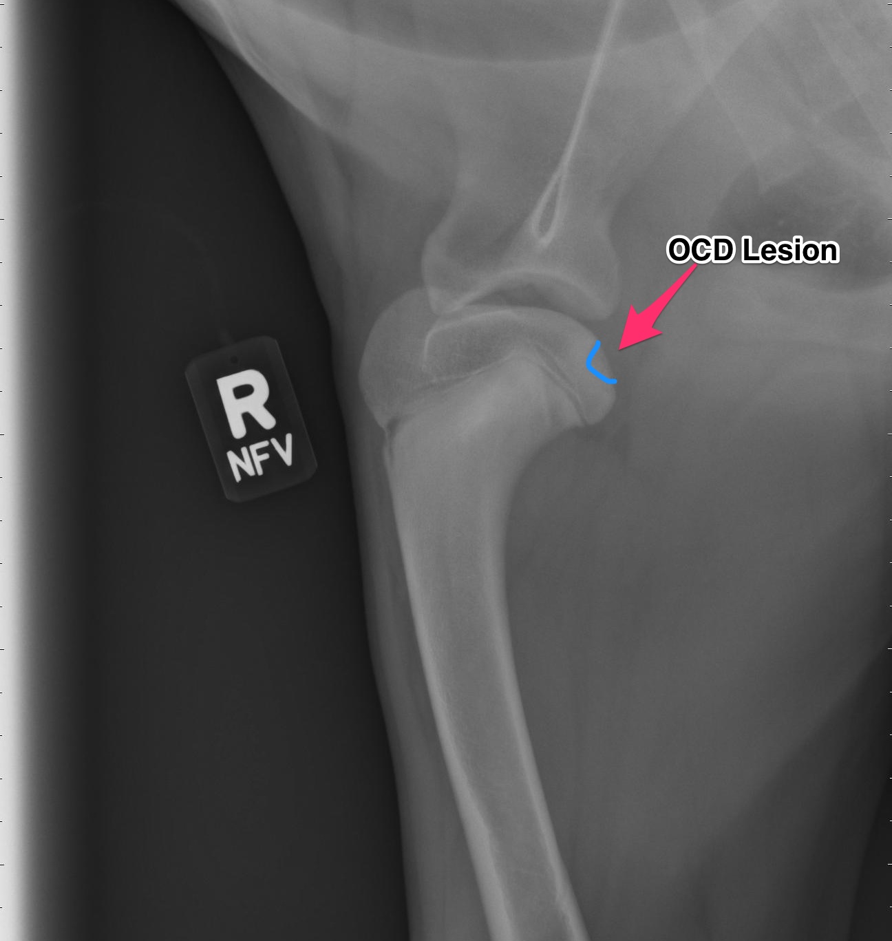
Radiographs (X-rays)
Above are x-rays of a left and right shoulder affected with OCD lesions. These are on the same patient. The images labeled with the left (L) marker has a flattened region noted by the arrow which is characteristic of OCD. The image on the right has the area highlighted in blue. While the lesion doesn’t look big, it can definitely cause a lot of pain and discomfort.
Another way to diagnostically evaluate the joint is with a computed tomography (CT) scan. This will give more detail into the region of interest. Generally this is not needed, however indications for it may be to evaluate the elbows as well.
Treatment:
For the best possible outcome do not delay treatment! At this time, the gold standard approach is arthroscopic debridement (removal) of the fragmented cartilage and the surrounding diseased cartilage and subchondral bone. Curettage may allow the now vacant cartilage bed to fill in more quickly with what is called fibrocartilage. I likened the removal of the fragment to old wallpaper removal (very much oversimplified). Once the old wallpaper bubbles and tears, you need to remove all the damaged wallpaper in the periphery or else the wallpaper will continue to peel.
If the cartilage is an osteochondrosis (OC) lesion and has not fragmented (OCD) non-surgical treatments (activity restriction, dietary restriction, etc) may be attempted and successful. Unfortunately, if OCD has not occurred then the patient will not be limping and most of these dogs go undiagnosed. It is my belief that any dog exhibiting pain/lameness with the presence of a radiographic (x-ray) OCD lesion ,should have surgery. Surgery will benefit them both in the short term and the long term.
There are older techniques of opening the joint to get access to the cartilage flap, however the recovery time on this type of procedure is significantly longer than with arthroscopy. Also, potential complications are increased with an “open” technique than with arthroscopic techniques. Arthroscopy is a minimally invasive tool that allows us to both diagnose and treat this condition. Generally speaking the patient can walk on the surgery leg (even if both legs have surgery at the same time!) following an arthroscopic procedure. Generally 2-3 small ports are placed over the shoulder (2-4mm in length) and this allows us access to the joint and work within the joint.
Recovery and Rehabilitation:
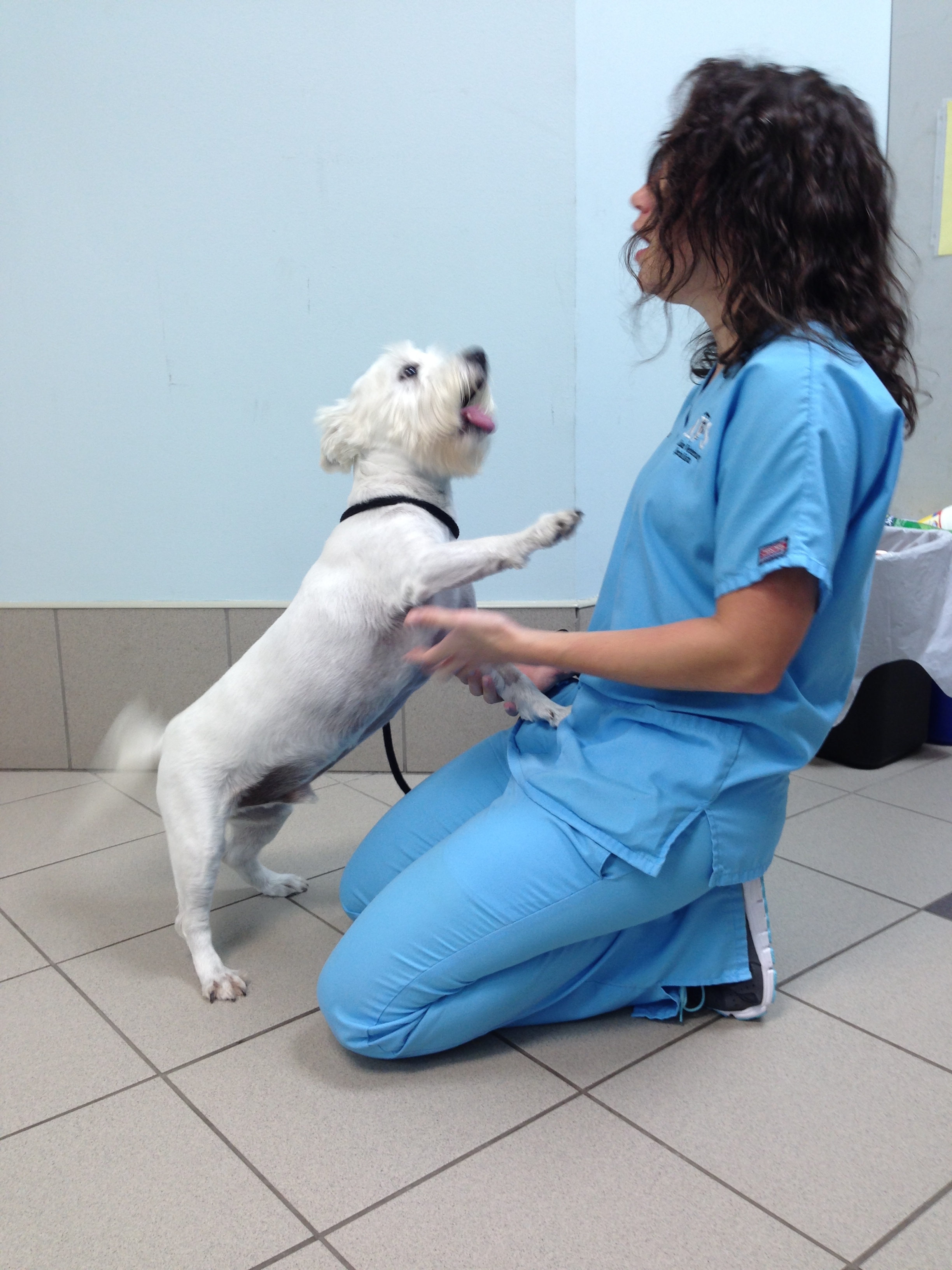
Recovery for the arthroscopic procedure is generally 4-6 weeks. Every surgeon has a different protocol for after surgery and I am very respectful of that. I prefer controlled movement for my patients. In the first two weeks, passive range of motion is very important, followed by active icing of the joint(s). Short leash based walks are started shortly after surgery and incrementally increased as we proceed through the recovery phase. Introduction into a formal rehabilitation program is recommended, however there are times when this is not possible and rehabilitation must be performed at home. Below is a patient that had a single shoulder arthroscopy, you can see how well they can walk following surgery (this is the following day)!
Prognosis:
When diagnosed and treated early, the dog affected with OCD can have a good prognosis and resume a normal or near normal activity level and quality of life. The longer the lesion is present, the more inflammation and arthritis will develop decreasing our success with surgery. Of the OCD lesions (shoulder, versus the other sites affected) this region has the best prognosis. I do encourage all my patients to continue on joint supplementation for life and to be removed from any breeding program.
Don’t forget to cast your vote for the PetPlan Vet of the Year!!! You can vote daily until October 17th!!! Show your support and click on the following link:
Tido two weeks after surgery!!! You can’t keep him down!!!
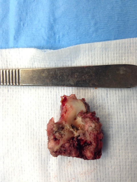
Meet Tido!!! Tido is a 6 1/2 year old West Highland White Terrier that came to us (Affiliated Veterinary Specialists – Orange Park) for a lower esophageal foreign body. He started showing signs of intermittent vomiting and regurgitation after swallowing his rawhide bone. Unfortunately, it became lodged in the portion of the esophagus that goes through his chest, just past his heart. Usually we can use a scope camera and remove the object without any incisions. The piece of rawhide was wedged in this area and was unable to be moved, so surgery was his only option.
This was the piece of rawhide that was lodged in Tido’s esophagus. It was nearly 4cm in length!!
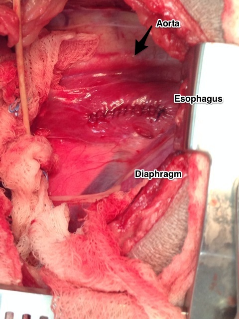
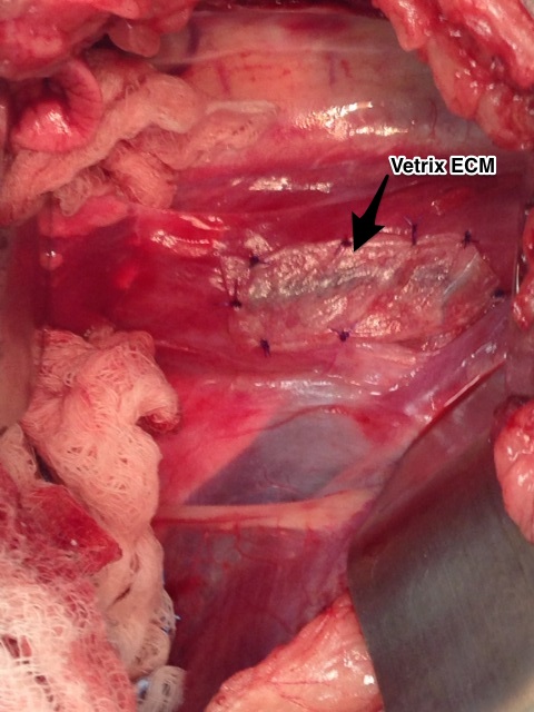
An incision was made in the chest and the large piece of rawhide was found in the esophagus just past the heart. An incision was made into the esophagus and the rawhide was removed. Surgery on the esophagus is a very delicate surgery. In this area we have big vessels (aorta) above the esophagus and the vena cava below. Just in front of the esophagus is the heart. Nearby, there are very important nerves (vagus) that course over the esophagus. Also, the esophagus has a harder time healing versus other areas of the gastrointestinal tract with a higher chance of stricture (narrowing due to scar tissue) formation.
View of the esophagus just past the heart.
After the rawhide was removed, the esophagus was closed in two layers and then a Vetrix Extracellular Matrix (ECM) sheet was placed. This will aid in healing by providing a scaffold for the tissue to heal and incorporate the bodies own stem cells to infiltrate the area. After the esophagus was closed, Tido’s chest was closed in a standard fashion.
Vetrix Extracellular Matrix placement over the esophageal incision.
Tido made an excellent recovery!!! He has been on a soft diet and no rawhides for him!!! In four weeks he should be able to resume his normal activity. At his two week recheck, you could never tell he had surgery. Way to go Tido!!!
Exciting news!!! Late last week I learned that I was nominated for the 2015 Petplan Pet Insurance (USA) Vet of the Year and that I was in the semifinals (6 out of 2300 nominations). I am honored and humbled by this nomination. If you click on the link (picture) below you can vote for the Vet of the Year (once daily). Every vote counts!!! Click on the following: