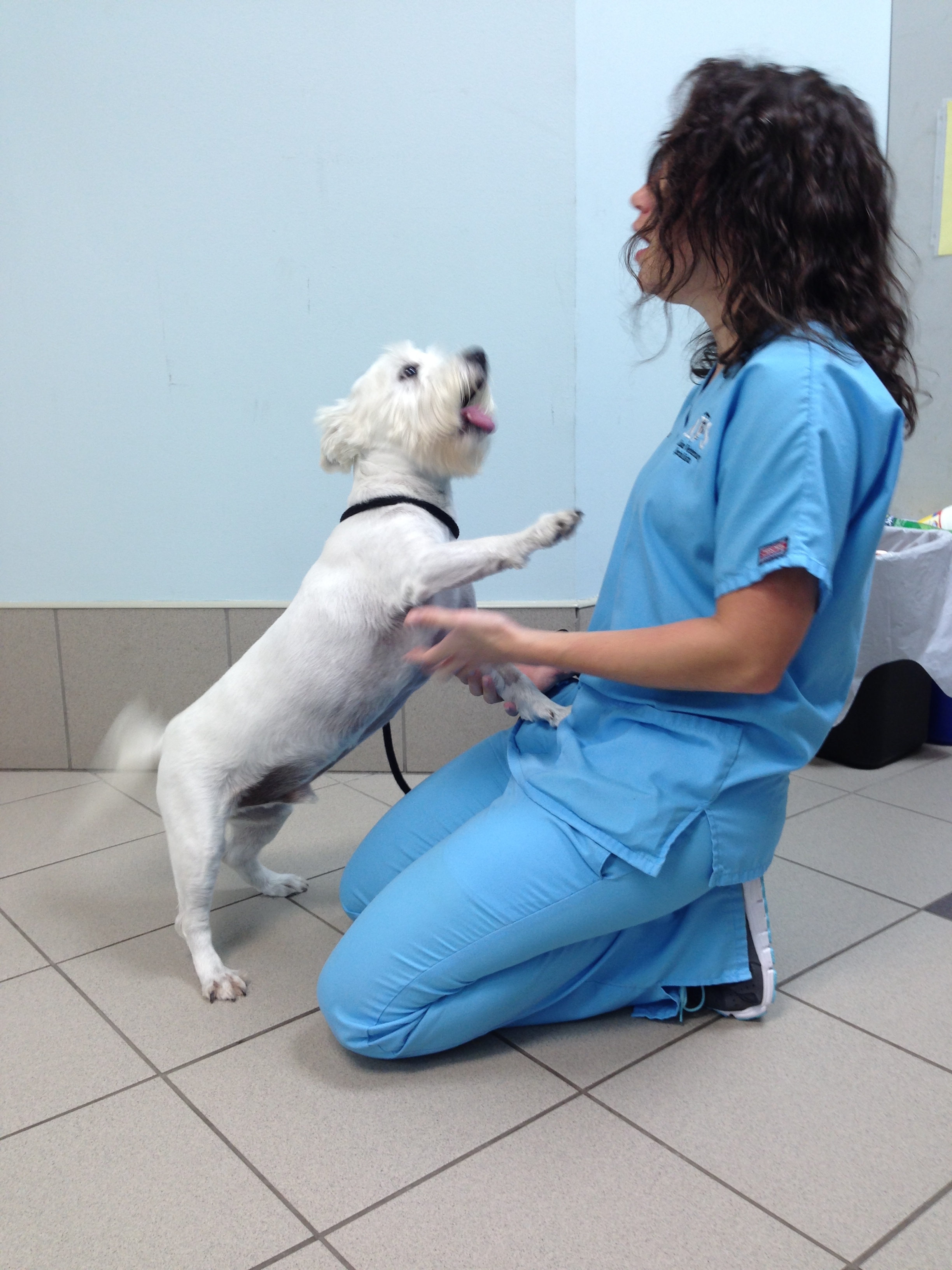
Tido two weeks after surgery!!! You can’t keep him down!!!
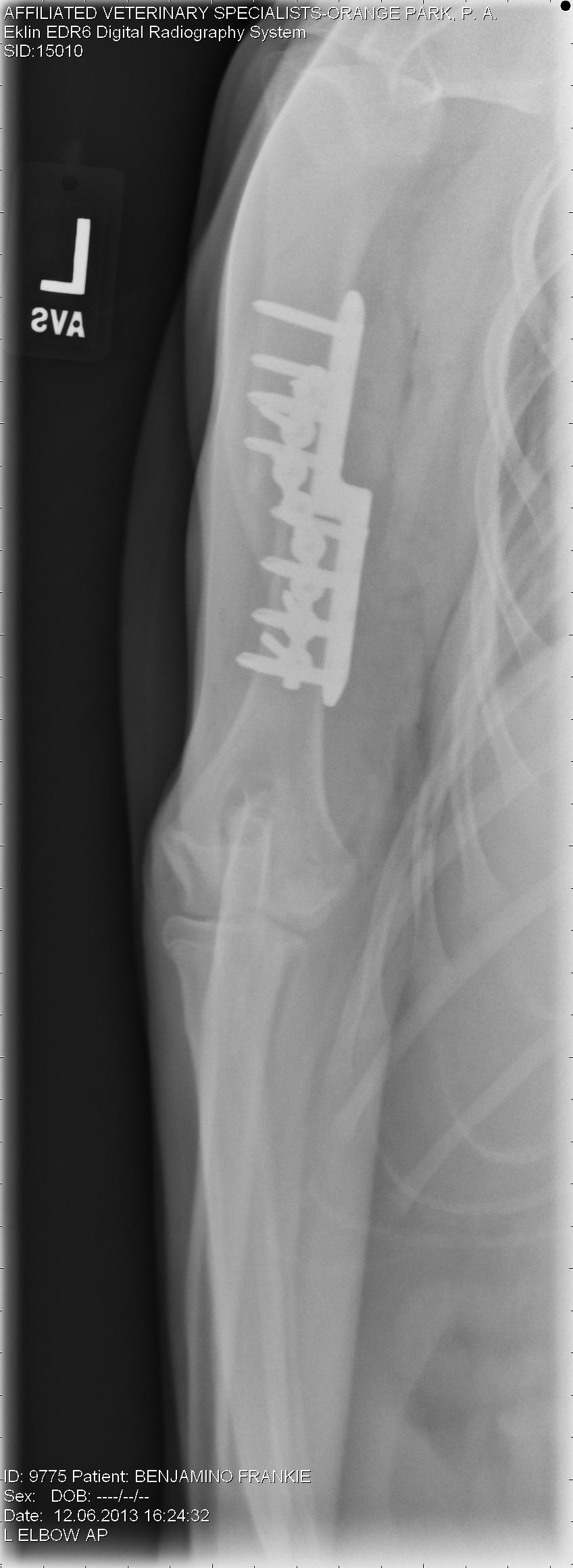
Meet Tido!!! Tido is a 6 1/2 year old West Highland White Terrier that came to us (Affiliated Veterinary Specialists – Orange Park) for a lower esophageal foreign body. He started showing signs of intermittent vomiting and regurgitation after swallowing his rawhide bone. Unfortunately, it became lodged in the portion of the esophagus that goes through his chest, just past his heart. Usually we can use a scope camera and remove the object without any incisions. The piece of rawhide was wedged in this area and was unable to be moved, so surgery was his only option.
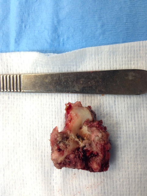
This was the piece of rawhide that was lodged in Tido’s esophagus. It was nearly 4cm in length!!
An incision was made in the chest and the large piece of rawhide was found in the esophagus just past the heart. An incision was made into the esophagus and the rawhide was removed. Surgery on the esophagus is a very delicate surgery. In this area we have big vessels (aorta) above the esophagus and the vena cava below. Just in front of the esophagus is the heart. Nearby, there are very important nerves (vagus) that course over the esophagus. Also, the esophagus has a harder time healing versus other areas of the gastrointestinal tract with a higher chance of stricture (narrowing due to scar tissue) formation.
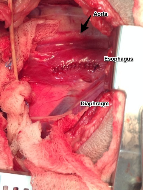
View of the esophagus just past the heart.
After the rawhide was removed, the esophagus was closed in two layers and then a Vetrix Extracellular Matrix (ECM) sheet was placed. This will aid in healing by providing a scaffold for the tissue to heal and incorporate the bodies own stem cells to infiltrate the area. After the esophagus was closed, Tido’s chest was closed in a standard fashion.
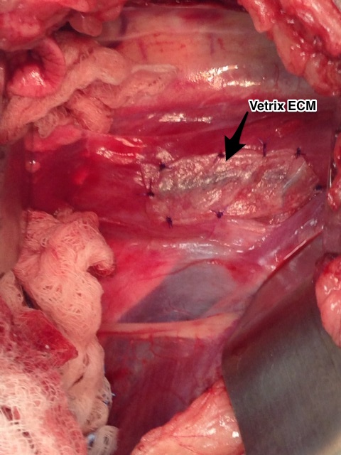
Vetrix Extracellular Matrix placement over the esophageal incision.
Tido made an excellent recovery!!! He has been on a soft diet and no rawhides for him!!! In four weeks he should be able to resume his normal activity. At his two week recheck, you could never tell he had surgery. Way to go Tido!!!