Frankie’s Journey: Part 1

Frankie and Vinnie relaxing.

Frankie and Vinnie relaxing.

Here are some of the diagnostics  that were performed on Frankie. As mentioned before, during his first visit a thorough examination was performed, which revealed congenital issues on all limbs. Also, when he presented he was overweight (for a 6 month old puppy). We do know that too rapid growth and obesity at a young age can preferentially express the genes responsible for these issues and exacerbate signs.

Here are his physical exam findings: lameness noted in all limb, more pronounced on his left side. Pain on hyperextension and hyperflexion of both elbows and pain on palpation of the medial (inside) compartment of both elbows. Pain on hyperextension and hyperflexion of both hips. Positive Ortolani tests on both hips – this is were the femoral head portion of the hip can be pushed out of the acetabulum (socket) and then falls back in; I have downloaded a video displaying this on Frankie.

Our first steps in diagnosis was to perform radiographs (x-rays) and a CT scan of the elbows. The x-rays confirmed both elbow dysplasia and hip dysplasia. Interestingly, Frankie had bilateral Osteochondritis Dissecans lesions, which isn’t commonly seen, but can be a part of elbow dysplasia.

The CT scan demonstrates the same lesions as the x-rays, but gives a different view. A CT scan is a very sensitive diagnostic test for elbow pathology, especially in cases that have not developed normal x-rays changes yet.

Left Elbow: Osteochondritis Dissecans (OCD).

Left Elbow: Osteochondritis Dissecans (OCD).

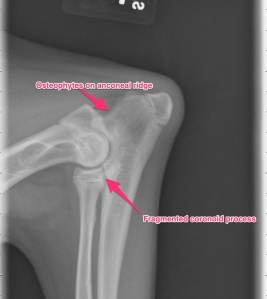
Lateral view of the left elbow: elbow dysplasia

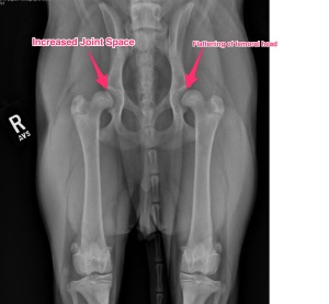
Bilateral Hip Dysplasia

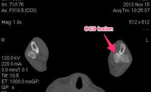
OCD lesion seen on the humerus.

Fragmented medial coronoid process.

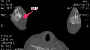
Fragmented medial coronoid process (FCP).

Brachycephalic Upper Airway Syndrome (BUAS) – physical exam

Most commonly the history of patients with BUAS are very similar. Generally, owners notice snoring and gradual progression of inspiratory stridor. Many times this will occur while the patient is a puppy and continue into adulthood. Other signs that are noted are increasing frequency of dyspnea especially during exercise or a hot environment. Another sign to look for in addition to the other is vomiting and/or regurgitation. This can be a compounding problem as it predisposes the pet to aspiration pneumonia.

On physical exam one of the first things that can be directly visualized is the nares being stenotic. The observant examiner will also note an increased upper respiratory noise with inspiratory stridor on auscultation(listening with a stethoscope. Typically the rest of the general physical examination is within normal limits.

Prior to performing a sedated oral exam various differentials should be on the list and should include neoplasia (oral/pharyngeal masses/cancer), tracheal collapse, laryngeal paralysis, lower airway disease and cardiac(heart)disease. Both lower airway diseases and cardiac disease can greatly exacerbate upper airway signs and if the patient is stable, these should be addressed first or at least concurrently.

Our next segment will be an overview of the surgical treatment and then we can wrap up this topic.

Happy Holidays

I want to take a moment to thank everyone for their continued interest and support of this blog. Your continued support has meant a lot to me and allowed me to continue to post various topics. I want to wish everyone Happy Holidays during this joyous time of the year! I wish you all the best in the New Year too! Keep your pets safe throughout this time, but definitely spoil them (we want them to ring in the New Years without any ailments)! Please continue to follow this blog!

Seasons Greetings!!

Kevin

Brachycephalic Obstructive Airway Syndrome (BOAS)

What makes up BOAS? The most common components of this disease are both stenotic nares(nostril) and an elongated soft palate. These two features are commonly seen. Other components are everted saccules, hypoplastic trachea, and secondary laryngeal collapse. In dogs that have stenotic nares the cartilage that makes up the nares is generally thicker and more condensed and oftentimes more medially displaced which causes an obstruction. Also, the conchae(cartilage in the nasal passageway) can be altered and displaced causing further airway turbulence and obstruction.

Anatomically the soft palate is just caudal(behind) to the hard palate and further divides the nasopharynx from the oropharynx (nasal passage from the mouth or oral cavity). In the normal patient the soft palate will typically extend caudally to just touch (slightly overlap) the epiglottis. In patients with an active obstruction this can be seen to extend 1-2 cm (or more) past the epiglottis. Due to this extension past the epiglottis, this tissue can actively obstruct airflow into the larynx and also become edematous(swollen) and undergo inflammation. Inflammation from this airway obstruction can extend to the tissue surround the pharyngeal region.

Generally considered to be a secondary side effect of the aforementioned conditions, everted saccules can develop due to the presence of increased air pressure of a prolonged period of time. Laryngeal saccules are normal out-pouchings noted adjacent to the vocal folds. Normally they extend outward away from the airway. After being under constant negative pressure these out-pouchings will evert and extend into the caudal laryngeal lumen causing an obstruction. Another secondary effect noted due to the constant high pressure is laryngeal collapse. This generally occurs in later stages of the disease process and will progress in severity. There is a grading scale that is used to assess the condition. Grade I laryngeal collapse is present when the laryngeal lumen is narrowed by everted saccules. Grade II collapse is characterized by both everted saccules and the cuneiform processes begin to collapse inward and fail to abduct during inspiration. Grade III collapse is characterized by the addition of the corniculate processes inwardly folding during inspiration, which signifies complete collapse.

The next posting will go over some of the physical exam findings and what we can do to help these patients out for the long run.

Note the narrow nostrils.

Note the narrow nostrils.

Treatment for an Intestinal Obstruction

Sorry about the nearly one month lapse in writing. I think it would be good to finish the topic on intestinal obstructions. The biggest question to be asked is, once the diagnosis is made – how do we correct the issue. The answer is one of three: 1. endoscopic removal of gastric foreign material, 2. surgical removal by an abdominal exploratory (or laparoscopy – only if a focal obstruction), and 3. hospitalization and fluids, if and only if , the foreign material is diagnosed as being in the large intestine/colon.

Generally, surgical explore of the abdomen and removal via a gastrotomy, enterotomy, or intestinal resection and anastomosis is recommended. Rather than go into the technical aspects of the surgeries, it would be better to put the emphasis of the importance of early detection and treatment of an intestinal obstruction, especially a complete obstruction. The biggest worry is both where in the gastrointestinal tract and to what extent did the material cause damage to the intestine. This can range from irritation and inflammation to perforation and necrosis (death) of the affected intestine. Early intervention is key in trying to reduce the risk of extensive damage. The more aggressive the procedure required increases post-operative risk to the patient.

Most patients will do well with surgery and recover uneventfully. Standard enterotomies and resection/anastomosis have a 10-15% complication rate when performed according to the literature. The most concerning complication is termed dehiscence (leaking of the intestinal suture line) and require another surgery to repair the area. Unfortunately, with more surgery required, the complication rate increases.

The best word of advice would be to attempt to identify possible foreign bodies and remove them them your pets reach. Some examples would be torn pieces of toys, squeakers from toys that have been removed, string/fishing line from cats, etc. However, if you do find your pets have the common signs – seek veterinary help right away.

An x-ray of a foreign body both in the stomach and within the intestines.

Minimally Invasive Surgery – Thoracoscopy

Thoracoscopy is the use of a rigid scope to evaluate the thoracic cavity. Typically this is performed in either dorsal recumbency (on their back) or lateral recumbency (on side) and involves an average of 3 portals. These patients typically experience a marked decrease in pain post-operatively when compared to a lateral thoracotomy or median sternotomy (open chest procedures).

Indications for thoracoscopy are as follows (not limited to): exploration of the thoracic cavity, pleural effusion of unknown origin, idiopathic pericardial effusion, lung resection, vascular ring anomaly, biopsies of the chest cavity, lymph node biopsy, pyothorax evaluation, etc. When appropriate, the major advantage is patient comfort, decreased morbidity, and in many cases better visualization. One of the most common reasons for thoracoscopy is pericardial effusion. This can be idiopathic, neoplastic or inflammatory. Therapy for pericardial effusions could include a pericardial window which can be done via thoracoscopy. Common risks with this procedure are as follows: herniation of the heart, fibrosis of the window, and continued hemorrhage.

Results of thoracoscopic pericardial windows are usually very successful when performed in the appropriate patient. It can provide long-term resolution in patients with idiopathic effusion and inflammatory disease. Quality of life can be improved in cases with cancer.

Overall, thoracoscopic procedures are increasing in popularity. The thought of being able to address surgical problems with minimal incision versus using large invasive incisions is very attractive. Please feel free to discuss these options with your veterinarian or veterinary surgeon.

Pericardial window being performed with Ligasure.

Minimally Invasive Surgery – 1

I hope everyone is having a great weekend! The next series of topics is join got focus on minimally invasive procedures. This is of particular interest of mine. There are multiple implications of this terms, I am going to focus on laparascopy, thoracoscopy and arthroscopy.

As medicine advances in both the human and veterinary world, we are starting to do more with less. What I mean is that through smaller incisions and better technology we address more and more surgical problems. The benefit of this is shorter hospital stays, decreased complication rte, decreased infection rates, and decreased soft tissue trauma and post-surgical pain. We are very lucky to have all this new technology available to us.

Here is a definition of the terms:

Laparascopy: Abdominal surgery being performed with a scope/camera via a small portal incision. Multiple other portals are made to introduce instruments. All procedures are performed within the abdominal cavity. Laparoscopic-assisted means that part of the procedure is done with cameras within the body cavity and part is done via small incisions outside of the abdomen.

Thoracoscopy: Same definition as above, however this pertains to the chest or thorax. This is a very useful modality and is used to treat various cardiac, pulmonary (lung) and esophageal problems.

Arthroscopy: Same definition as above, however this is performed on the joints. This is a very common use in veterinary medicine and is thought to be the gold standard in joint evaluation. The most common joints evaluated through this are the elbows, shoulders and knees, however the wrists (carpi), ankles (hocks/tarsi), and hips can be evaluated. Just think, if you have an ACL injury, it is very common for you to have your knee scoped, why not your best friend?