
Welcome to “A Veterinarian’s Perspective”. Thank you for taking the time to visit this site. One of the many goals is to give accurate and useful information about a wide array of veterinary medicine topics. There are many places online one can obtain information; some “good” and some “not so good”. This site is geared to both the veterinary professional and the non-veterinary trained. Another passion and new facet to this website/blog is in the realm of business management as it pertains to the practice of veterinary medicine. Look for some fresh, useful topics in the near future. By all means, feel free to give suggestions for topics!
cats
Top 5 List for a Safer Holiday Season
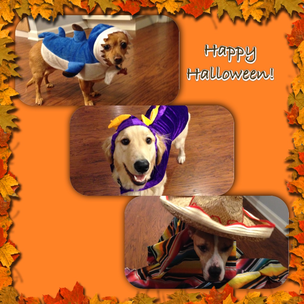
As Halloween and the upcoming holidays are rapidly approaching, we are often wrapped up in family gathering, parties, and other activities and forget about the well being of our beloved pets. I tried compiling a short list to help keep our pets healthy and out of trouble this holiday season. This should ensure that everyone has a happy holiday season and may save you and your pet from needing emergency trips to veterinarian or emergency clinic.
1. Keep candy away from pets, in particular chocolate and candies made with xylitol and other sweeteners. While these taste good, they can have very harmful effects on your pet, ranging from liver failure, seizures, and as severe as death. If you suspect your pet has consumed any of these, seek veterinary care immediately.
2. Keep a close on the whereabouts of your pets. With all the excitement and increased visitors during this time, make sure your pets are accounted for and haven’t run off. Missing pets and subsequent trauma, such as being hit by a vehicle is an all to common occurrence during this time. Make sure your pets are in a safe place when company is over.
3. Keep pets out of the garbage and from grabbing food off the table. Bones and fatty meats can cause illness in our pets, especially dogs. Bones can cause a lot of irritation and in some cases puncture the gastrointestinal tracts. Fatty foods are not good for our pets and can cause pancreatitis and other gastrointestinal issues. Pancreatitis can range in severity and needs to be treated by your veterinarian.
4. Keep cords and electrical wiring away from your pets. Both cats and dogs can find wires enticing. Electrocution injury can be very severe and cause death in some cases. If you believe your pet to have be electrocuted, have them evaluated by your veterinarian immediately.
5. Keep easily ingestible objects away from your pets. Objects that can be easily swallowed can cause gastrointestinal irritation and obstruction. Some objects that can become obstructive are clothes, small toys, tinsel, etc. Gastrointestinal obstructions demand immediate veterinary care. There are times when the object can pass, but most of the time your pet will need surgery to relieve the obstruction, Surgery can range from a single incision in the stomach to removal of a segment of intestine. In extreme cases this condition can be fatal.
Golden Retrievers Provide Comfort for Citizens of Newtown
Golden Retrievers Provide Comfort for Citizens of Newtown.
This is such a touching story, so I choose to share this as it is animal related. It really demonstrates the bond that we have with our pets and how much influence they really have!!
Brachycephalic Upper Airway Syndrome (BUAS) – physical exam
Most commonly the history of patients with BUAS are very similar. Generally, owners notice snoring and gradual progression of inspiratory stridor. Many times this will occur while the patient is a puppy and continue into adulthood. Other signs that are noted are increasing frequency of dyspnea especially during exercise or a hot environment. Another sign to look for in addition to the other is vomiting and/or regurgitation. This can be a compounding problem as it predisposes the pet to aspiration pneumonia.
On physical exam one of the first things that can be directly visualized is the nares being stenotic. The observant examiner will also note an increased upper respiratory noise with inspiratory stridor on auscultation(listening with a stethoscope. Typically the rest of the general physical examination is within normal limits.
Prior to performing a sedated oral exam various differentials should be on the list and should include neoplasia (oral/pharyngeal masses/cancer), tracheal collapse, laryngeal paralysis, lower airway disease and cardiac(heart)disease. Both lower airway diseases and cardiac disease can greatly exacerbate upper airway signs and if the patient is stable, these should be addressed first or at least concurrently.
Our next segment will be an overview of the surgical treatment and then we can wrap up this topic.
Happy Holidays
I want to take a moment to thank everyone for their continued interest and support of this blog. Your continued support has meant a lot to me and allowed me to continue to post various topics. I want to wish everyone Happy Holidays during this joyous time of the year! I wish you all the best in the New Year too! Keep your pets safe throughout this time, but definitely spoil them (we want them to ring in the New Years without any ailments)! Please continue to follow this blog!
Seasons Greetings!!
Kevin
Brachycephalic Obstructive Airway Syndrome (BOAS)
What makes up BOAS? The most common components of this disease are both stenotic nares(nostril) and an elongated soft palate. These two features are commonly seen. Other components are everted saccules, hypoplastic trachea, and secondary laryngeal collapse. In dogs that have stenotic nares the cartilage that makes up the nares is generally thicker and more condensed and oftentimes more medially displaced which causes an obstruction. Also, the conchae(cartilage in the nasal passageway) can be altered and displaced causing further airway turbulence and obstruction.
Anatomically the soft palate is just caudal(behind) to the hard palate and further divides the nasopharynx from the oropharynx (nasal passage from the mouth or oral cavity). In the normal patient the soft palate will typically extend caudally to just touch (slightly overlap) the epiglottis. In patients with an active obstruction this can be seen to extend 1-2 cm (or more) past the epiglottis. Due to this extension past the epiglottis, this tissue can actively obstruct airflow into the larynx and also become edematous(swollen) and undergo inflammation. Inflammation from this airway obstruction can extend to the tissue surround the pharyngeal region.
Generally considered to be a secondary side effect of the aforementioned conditions, everted saccules can develop due to the presence of increased air pressure of a prolonged period of time. Laryngeal saccules are normal out-pouchings noted adjacent to the vocal folds. Normally they extend outward away from the airway. After being under constant negative pressure these out-pouchings will evert and extend into the caudal laryngeal lumen causing an obstruction. Another secondary effect noted due to the constant high pressure is laryngeal collapse. This generally occurs in later stages of the disease process and will progress in severity. There is a grading scale that is used to assess the condition. Grade I laryngeal collapse is present when the laryngeal lumen is narrowed by everted saccules. Grade II collapse is characterized by both everted saccules and the cuneiform processes begin to collapse inward and fail to abduct during inspiration. Grade III collapse is characterized by the addition of the corniculate processes inwardly folding during inspiration, which signifies complete collapse.
The next posting will go over some of the physical exam findings and what we can do to help these patients out for the long run.

Note the narrow nostrils.
Upper Respiratory Issues – Brachycephalic Upper Airway Syndrome (BUAS)
Overview:
A very common airway problem seen in small dogs and some bigger dogs is a syndrome termed Brachycephalic Upper Airway Syndrome (BUAS). This disease process affects brachycephalic dogs due to the development of their skull structure. These dogs can be thought of having the same anatomy as a longer nose dog, just in a much smaller area. As one could image, this can cause difficulty in the passage of air as it traverses through the nasal passage and into the naso- and oropharynx causing an increased pressure.
Common breeds that are affected and grouped into the breed type(brachycephalic) range from the Bulldog to the Cavalier King Charles Spaniels. Dogs that are affected by BUAS can present with many different symptoms that all involve the passage of air and include snoring, stridor, exercise intolerance, apnea and even gastrointestinal side effects such as vomiting and regurgitation. There are factors that can worsen the signs such as weight gain, allergies, environment conditions etc. Factors that can be controlled should be addressed.
In the next segment we will look at the physical features of dogs that have brachycephalic upper airway syndrome. Feel free to ask questions.
Pug and stenotic nares – after surgery
Treatment for an Intestinal Obstruction
Sorry about the nearly one month lapse in writing. I think it would be good to finish the topic on intestinal obstructions. The biggest question to be asked is, once the diagnosis is made – how do we correct the issue. The answer is one of three: 1. endoscopic removal of gastric foreign material, 2. surgical removal by an abdominal exploratory (or laparoscopy – only if a focal obstruction), and 3. hospitalization and fluids, if and only if , the foreign material is diagnosed as being in the large intestine/colon.
Generally, surgical explore of the abdomen and removal via a gastrotomy, enterotomy, or intestinal resection and anastomosis is recommended. Rather than go into the technical aspects of the surgeries, it would be better to put the emphasis of the importance of early detection and treatment of an intestinal obstruction, especially a complete obstruction. The biggest worry is both where in the gastrointestinal tract and to what extent did the material cause damage to the intestine. This can range from irritation and inflammation to perforation and necrosis (death) of the affected intestine. Early intervention is key in trying to reduce the risk of extensive damage. The more aggressive the procedure required increases post-operative risk to the patient.
Most patients will do well with surgery and recover uneventfully. Standard enterotomies and resection/anastomosis have a 10-15% complication rate when performed according to the literature. The most concerning complication is termed dehiscence (leaking of the intestinal suture line) and require another surgery to repair the area. Unfortunately, with more surgery required, the complication rate increases.
The best word of advice would be to attempt to identify possible foreign bodies and remove them them your pets reach. Some examples would be torn pieces of toys, squeakers from toys that have been removed, string/fishing line from cats, etc. However, if you do find your pets have the common signs – seek veterinary help right away.

An x-ray of a foreign body both in the stomach and within the intestines.
Common Signs with Intestinal Obstructions
Welcome back. This shouldn’t take too long, but let’s review some of the more common signs seen with intestinal obstructions. The most common sign would be vomiting and generally not a one time occurrence. This will usually be profuse vomiting (but can vary with every patient). Other signs to look for is anorexia (not wanting to eat), lethargy, and abdominal pain. As with any type of foreign body the gastrointestinal tract can become perforated and significantly worse signs can become evident. Whenever an intestinal foreign body is suspected immediate veterinary care (whether your primary veterinarian or an emergency clinic) is highly recommended. Generally these signs will occur very acutely (all of a sudden) once the object begins to obstruct the intestine. We don’t always know what our dogs get into, especially if they are left unattended or go outside in the back and unsupervised. It seems like many owner don’t know what there pet got into and swallowed.
During the initial evaluation, your veterinarian may recommend some diagnostic tests to help support the diagnosis of an intestinal foreign body and justify surgery. Most of the time the physical exam will show, dehydration (high heart rate, dry mucus membranes, etc), evidence of vomiting, pain on abdominal palpation, fever (if perforated intestines) and other various signs. Some dogs when presented early show few signs and are very stable, other dogs show very severe signs and may be very unstable and require aggressive supportive care including aggressive fluid management and other treatments.
The standard diagnostics after initial triage (physical exam, blood pressure, etc) typically include abdominal radiographs (x-rays), complete blood work, possible chest radiographs (if aspiration suspect or in the geriatric dog) and possible abdominal ultrasound if the radiographs are non-diagnostic. Some may also recommend contrast radiographs with barium contrast in an attempt to highlight the foreign body.
Once a presumptive diagnosis is made and the patient is deemed stable surgery generally is the next step. If the foreign body is only in the stomach, endoscopy may be performed succesfully to remove the object.
Minimally Invasive Surgery – Thoracoscopy
Thoracoscopy is the use of a rigid scope to evaluate the thoracic cavity. Typically this is performed in either dorsal recumbency (on their back) or lateral recumbency (on side) and involves an average of 3 portals. These patients typically experience a marked decrease in pain post-operatively when compared to a lateral thoracotomy or median sternotomy (open chest procedures).
Indications for thoracoscopy are as follows (not limited to): exploration of the thoracic cavity, pleural effusion of unknown origin, idiopathic pericardial effusion, lung resection, vascular ring anomaly, biopsies of the chest cavity, lymph node biopsy, pyothorax evaluation, etc. When appropriate, the major advantage is patient comfort, decreased morbidity, and in many cases better visualization. One of the most common reasons for thoracoscopy is pericardial effusion. This can be idiopathic, neoplastic or inflammatory. Therapy for pericardial effusions could include a pericardial window which can be done via thoracoscopy. Common risks with this procedure are as follows: herniation of the heart, fibrosis of the window, and continued hemorrhage.
Results of thoracoscopic pericardial windows are usually very successful when performed in the appropriate patient. It can provide long-term resolution in patients with idiopathic effusion and inflammatory disease. Quality of life can be improved in cases with cancer.
Overall, thoracoscopic procedures are increasing in popularity. The thought of being able to address surgical problems with minimal incision versus using large invasive incisions is very attractive. Please feel free to discuss these options with your veterinarian or veterinary surgeon.

Pericardial window being performed with Ligasure.
