Laryngeal Paralysis

Laryngeal Paralysis

cropped-8135862891_b252936d62_b_4.jpg

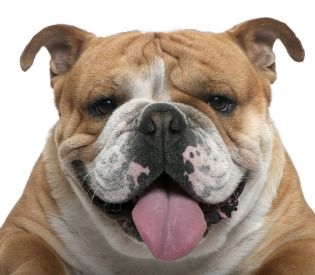
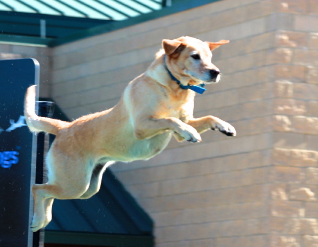
Spring and summer bring about so many good things: the beach, warm weather, family gatherings, to name a few; and then some bad things: bugs, sweltering heat and humidity, allergies, and the list goes on. For your pets, especially your older retrievers and short nosed (brachycephalic) breeds like the pug and bulldog, the heat and humidity can spell danger due to airway conditions. For more information on the brachycephalic dog breathing issues, see my other post  http://wp.me/p2vvxS-2R . This article will focus on a condition called laryngeal paralysis which typically affects our large breed dogs, such as the Labrador Retriever and similar breeds, although it has been seen in cats (rarely) and is a defined disease process in horses.

What is laryngeal paralysis?

Laryngeal paralysis can be as bad as it sounds. The larynx is at the back of the mouth and allows the passage of air into the windpipe (trachea). In the video below, it mimics swinging doors and the cartilages (arytenoid cartilage) that form the larynx will open when breathing “in” (inspiration) and open when breathing “out” (expiration). It remains closed during other actions, like eating and swallowing, thus stopping food, water, saliva from going down the trachea. There is a muscle that controls the opening of these cartilages. The muscle (cricoarytenoideus dorsalis muscle) sits on top of the cartilages on both sides and actively contracts to open the cartilages during inspiration. The opening of the cartilages when exhaling is passive as the air blows open the cartilages. Laryngeal paralysis is a condition where the nerves that feed this muscle are not working properly and the muscle atrophies and is nonfunctional – hence the larynx is paralyzed and can’t move normally.

What causes this condition???

In most dogs, we do not know the reason for this condition. We divide the condition into two general types: 1. congenital and 2. acquired. In congenital, this condition is usually seen at an early age and is thought to be hereditary. Some common breeds affected are Siberian Huskies, Bulldogs, Rottweilers, etc. In the second form (acquired), it simply means that the disease occurs secondary from other issues. When we think of causes we have to ask ourselves, what can cause damage/changes to the nerve (recurrent laryngeal nerve) that feeds the cricoarytenoideus dorsalis muscle? Conditions that we evaluate for typically are as follows: cervical (neck) tumors, chest/lung tumors, myasthenia gravis, peripheral neuropathies, previous neck (cervical) trauma, and endocrine diseases. Most of the time, we do not find a direct cause and suspect an undiagnosed peripheral neuropathy as the underlying cause. When we do not know the actual cause we term the disease “idiopathic”. Some recent studies (Stanley, et al) have shown that most (if not all) patients with idiopathic laryngeal paralysis will begin to display some generalized neurologic signs within 1-2 years following the diagnosis.

What are the signs of acquired laryngeal paralysis???

Typically, this affects our larger breed dogs, with the Labrador Retriever being the poster child for this disease. The dogs affected are generally middle to older in age, and either male or female. The most common signs noticed is difficulty breathing, especially when exercising or excited and gagging/coughing when eating/drinking. This is a progressive disease, so signs usually begin with mild changes and become more severe, which can be over months to years. You may also notice a change in the pitch of your dogs bark (voice). Most of the time, we can arrive at a presumptive diagnosis just listening to your pet breathing. As the disease progresses, the affected dog becomes more at risk, and can have a respiratory emergency if not managed appropriately, which can be fatal. Below is a video (the audio is most important) of a dog with laryngeal paralysis:

What diagnostics are involved with laryngeal paralysis???

To begin, a thorough general and neurologic examination is needed for your pet. At minimum, a complete blood work, including a thyroid screening panel, and chest (thoracic) x-rays are needed. The importance of these is to look for other disease processes that may be going on and to ensure that the major organs are functioning appropriately. Why the thyroid panel? Hyopthyroidism (low thyroid hormone production) can cause various neuromuscular issues. With the chest x-rays we are looking for any masses, changes to the esophagus size (megaesophagus) and signs of aspiration pneumonia, which can be seen secondary with laryngeal paralysis. Because most of the patients I see with this condition are older and we are assessing for surgery, I highly recommend an abdominal ultrasound by an experienced ultrasonographer to look for any other concurrent diseases. Bear in mind, if your pet is in a respiratory crisis some of these steps may be done out of order to adequately stabilize the patient.

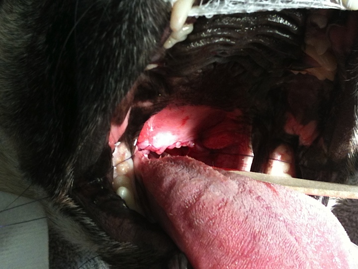
The best way to confirm the suspected diagnosis of laryngeal paralysis is to look directly at the larynx and assess the functioning of the laryngeal cartilages. This is typically done by inducing a light plane of anesthesia and looking at the back of the mouth. The proper assessment is sort of an art and takes practice to be comfortable making the diagnosis. In addtion to evaluating the larynx, time is taken to look at the rest of the oral cavity for other potential causes. As a surgeon, my preference is to do this examination directly prior to surgery to minimize the amount times the pet needs to undergo anesthesia.

Below is a video demonstrating laryngeal paralysis. The laryngeal opening can be seen and you will notice that it is not moving much at all during the phases of breathing.

How can I treat my pet once laryngeal paralysis is diagnosed???

Probably the better questions is when do I treat? Once a diagnosis is made, then a decision needs to be made. Since this is a progressive disease, if only one side of the larynx is affected then surgical options will most likely be delayed. The most typical treatment for idiopathic laryngeal paralysis is surgical. To date, there is no medical therapy that will restore the function of the larynx. Conservative management will typically incorporate ways to keep your pet cool (air conditioned environment), sedation possibly, and decreasing environmental allergens. If, during our pre surgical diagnostics, we find other issues, changes may be made to the treatment plan. There are some findings that may make your veterinarian reconsider your pet being a good surgical candidate, such as an enlarged esophagus (megaesophagus). The main reason to proceed forward with surgery is to improve your pets quality of life for however long that may be, as well as improve your (as the owner) life by providing more quality time together. There are risks both with surgery and without surgery.

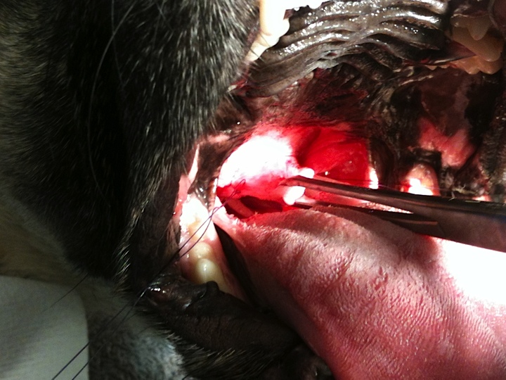
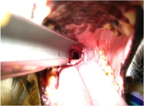
The standard procedure to open the airway is called an arytenoid lateralization (laryngeal tie-back). This is a procedure that pulls one side of the laryngeal cartilages back, permanently opening one side of the larynx. In effect, we override the normal muscular action of the larynx. We gain access to the larynx by an incision made on the side of the neck. None of the work is done within the mouth. There are other procedures that remove the arytenoid cartilage portion of the larynx to permanently open the larynx from within the mouth, called an arytenoidectomy. This procedure, in my opinion, has not been evaluated as much as the “tie-back” procedure.

Below is a picture of an arytenoid lateralization. Notice the difference on the opening from the previous video.

Tie back

What are the risks with and without surgery and what is the typical outcome?

No procedure is without inherent risks, unfortunately. The risks and benefits of any procedure must always be weighed and discussed with your veterinarian and veterinary surgeon. The most common post-operative complication is aspiration pneumonia. Recent literature cites about a 12-15% risk of aspiration pneumonia following surgery, with the most critical time period being the actual recovery from surgery and the immediate post-operative period. Some medications can be administered that help reduce vomiting, regurgitation and increasing the tone of the lower esophageal sphincter muscle – all aimed at lowering this risk. Most of the time aspiration pneumonia, if caught early, can be treated successfully with antibiotic therapy and supportive care (depending on severity). In a small number of patients, aspiration pneumonia can be fatal. Other complications are break down of the “tie-back” suture and incisional complications such as seroma and abscess/infection. Anesthesia complications can arise with any anesthesia/surgical event, however with proper screening, this risk can be minimized. My feeling is that even dogs prior to a “tie-back” procedure have a higher risk of aspiration pneumonia because the protective mechanism of the larynx is not functioning properly.

Surgically addressing this condition can be life saving and drastically improve the quality of your pets life. Most owners (~90%) are happy they made the decision to proceed forward with surgery and are pleased with the improved quality of life for their pet. If you notice any of these changes to your pet, please plan to see your veterinarian to see if they are a candidate for surgery. While the above article is long, it does not include everything related to this disease, if you have questions, just ask!!!

Kevin Benjamino, DVM, DACVS

Copyright  2015

Pets and Decorations Don’t Mix

A timely discussion about some of the hazards that may be around the home during the holiday season.

Brachycephalic Obstructive Airway Syndrome – Treatment

Elongated soft palate

Elongated soft palate

Shortened soft palate

Shortened soft palate

The mainstay of treatment for BOAS remains surgical, however some medical measures can also be taken. When considering medical management, it is important to focus on factors that can cause worsening of the signs, such as weight loss and allergies. Other factors to consider are housing the patient in a cool environment, avoiding the use of neck leads, decreased activity levels and the use of gastroprotectants for any concurrent vomiting or regurgitation. Typically medical management is used after (and in conjunction with) surgical management.

There are many questions that arise when considering surgical management and one of the biggest is when do you consider surgery? BOAS can be seen even in puppies and it is recommended that an evaluation be performed in dogs that are predisposed to this condition. Early management can halt or delay the progression that is typically seen, especially laryngeal collapse.

There are various methods to widen the nares(nostrils). The most common technique is the vertical wedge resection, where a wedge of tissue is removed with the apex of the triangle at the dorsal surface. An absorbable suture can be placed to control bleeding. It is important to make sure that the nares is wide enough to increase airflow.

An elongated soft palate is one of the most common features of BOAS. The assessment and skilled resection is key. If too much is removed, then there is a communication between the oropharynx and nasopharynx. If not enough is removed then the problem still exists.  The most common technique employed is resection with metzenbaum scissors and suture. Sharp excision of the soft palate generally ensures the least amount of inflammation. Other methods, such as CO2 laser and Ligasure, have been described and can be successful. Complications that can arise are as follows: bleeding, inflammation, chronic granulation tissue formation, and further elongation of the soft palate over time.

As mentioned previously, the presence of everted saccules characterizes the patient with stage I laryngeal collapse. There is some controversy as to whether or not everted saccules should be addressed surgically.

With patients that have grade II and III laryngeal collapse surgical correction is more difficult. When collapse is present it is always recommend to correct what is correctable, however the larynx will never be functional again. Some propose modified laryngeal tieback procedures with mixed outcomes. A permanent tracheostomy becomes a very viable option. By performing a permanent tracheostomy the entire upper airway is by-passed.

When counseling owners, generally dogs affected with BOAS have a favorable prognosis. Success is solely dependent on progression of disease. Education of owners should start when the patient is a puppy to avoid worsening. When a patient progresses to laryngeal collapse prognosis decreases greatly, as well as hospitalization time.


Brachycephalic Upper Airway Syndrome (BUAS) – diagnosis

Diagnostic testing:

In addition to direct visualization of structures it is wise to evaluate both the neck and chest. This is most commonly performed with radiographs (X-rays). Structures to evaluate include the oro/nasopharynx, trachea, lungs (for pneumonia, etc), and cardiac disease. If cardiac disease is suspected due to auscultation and radiographs, an echocardiogram is recommended. A complete blood count (CBC), chemistry and urinalysis should be performed prior to anesthesia to assess total body function and if your pet is a good anesthetic and surgical candidate.

A very important part of the diagnostic workup performed just before surgery is the oral examination. One way to evaluate the upper airway is endoscopy of both the larynx and esophagus. This will provide a very thorough evaluation of the soft palate, ventricles and laryngeal function.

Direct visualization is the most common way of diagnosing the associated factors of the brachycephalic dog. The nares are narrower than a normal dog and the airflow is distorted.  For a sedated oral exam, the patient is typically anesthetized (light plane of anesthesia). Evaluating the laryngeal function in these dogs is very important in differentiating laryngeal paralysis from laryngeal collapse.

The mainstay of treatment for BUAS remains surgical, however some medical measures can also be taken. When considering medical management, it is important to focus on factors that can cause worsening of the signs, such as weight loss and allergies. Other factors to consider are housing the patient in a cool environment, avoiding the use of neck leads, decreased activity levels and the use of gastroprotectants for any concurrent vomiting or regurgitation.


Brachycephalic Upper Airway Syndrome (BUAS) – physical exam

Most commonly the history of patients with BUAS are very similar. Generally, owners notice snoring and gradual progression of inspiratory stridor. Many times this will occur while the patient is a puppy and continue into adulthood. Other signs that are noted are increasing frequency of dyspnea especially during exercise or a hot environment. Another sign to look for in addition to the other is vomiting and/or regurgitation. This can be a compounding problem as it predisposes the pet to aspiration pneumonia.

On physical exam one of the first things that can be directly visualized is the nares being stenotic. The observant examiner will also note an increased upper respiratory noise with inspiratory stridor on auscultation(listening with a stethoscope. Typically the rest of the general physical examination is within normal limits.

Prior to performing a sedated oral exam various differentials should be on the list and should include neoplasia (oral/pharyngeal masses/cancer), tracheal collapse, laryngeal paralysis, lower airway disease and cardiac(heart)disease. Both lower airway diseases and cardiac disease can greatly exacerbate upper airway signs and if the patient is stable, these should be addressed first or at least concurrently.

Our next segment will be an overview of the surgical treatment and then we can wrap up this topic.

Happy Holidays

I want to take a moment to thank everyone for their continued interest and support of this blog. Your continued support has meant a lot to me and allowed me to continue to post various topics. I want to wish everyone Happy Holidays during this joyous time of the year! I wish you all the best in the New Year too! Keep your pets safe throughout this time, but definitely spoil them (we want them to ring in the New Years without any ailments)! Please continue to follow this blog!

Seasons Greetings!!

Kevin

Brachycephalic Obstructive Airway Syndrome (BOAS)

What makes up BOAS? The most common components of this disease are both stenotic nares(nostril) and an elongated soft palate. These two features are commonly seen. Other components are everted saccules, hypoplastic trachea, and secondary laryngeal collapse. In dogs that have stenotic nares the cartilage that makes up the nares is generally thicker and more condensed and oftentimes more medially displaced which causes an obstruction. Also, the conchae(cartilage in the nasal passageway) can be altered and displaced causing further airway turbulence and obstruction.

Anatomically the soft palate is just caudal(behind) to the hard palate and further divides the nasopharynx from the oropharynx (nasal passage from the mouth or oral cavity). In the normal patient the soft palate will typically extend caudally to just touch (slightly overlap) the epiglottis. In patients with an active obstruction this can be seen to extend 1-2 cm (or more) past the epiglottis. Due to this extension past the epiglottis, this tissue can actively obstruct airflow into the larynx and also become edematous(swollen) and undergo inflammation. Inflammation from this airway obstruction can extend to the tissue surround the pharyngeal region.

Generally considered to be a secondary side effect of the aforementioned conditions, everted saccules can develop due to the presence of increased air pressure of a prolonged period of time. Laryngeal saccules are normal out-pouchings noted adjacent to the vocal folds. Normally they extend outward away from the airway. After being under constant negative pressure these out-pouchings will evert and extend into the caudal laryngeal lumen causing an obstruction. Another secondary effect noted due to the constant high pressure is laryngeal collapse. This generally occurs in later stages of the disease process and will progress in severity. There is a grading scale that is used to assess the condition. Grade I laryngeal collapse is present when the laryngeal lumen is narrowed by everted saccules. Grade II collapse is characterized by both everted saccules and the cuneiform processes begin to collapse inward and fail to abduct during inspiration. Grade III collapse is characterized by the addition of the corniculate processes inwardly folding during inspiration, which signifies complete collapse.

The next posting will go over some of the physical exam findings and what we can do to help these patients out for the long run.

Note the narrow nostrils.

Note the narrow nostrils.

Upper Respiratory Issues – Brachycephalic Upper Airway Syndrome (BUAS)

Overview:

A very common airway problem seen in small dogs and some bigger dogs is a syndrome termed Brachycephalic Upper Airway Syndrome (BUAS). This disease process affects brachycephalic dogs due to the development of their skull structure. These dogs can be thought of having the same anatomy as a longer nose dog, just in a much smaller area. As one could image, this can cause difficulty in the passage of air as it traverses through the nasal passage and into the naso- and oropharynx causing an increased pressure.

Common breeds that are affected and grouped into the breed type(brachycephalic) range from the Bulldog to the Cavalier King Charles Spaniels. Dogs that are affected by BUAS can present with many different symptoms that all involve the passage of air and include snoring, stridor, exercise intolerance, apnea and even gastrointestinal side effects such as vomiting and regurgitation. There are factors that can worsen the signs such as weight gain, allergies, environment conditions etc. Factors that can be controlled should be addressed.

In the next segment we will look at the physical features of dogs that have brachycephalic upper airway syndrome. Feel free to ask questions.

Pug and stenotic nares - after surgery

Pug and stenotic nares – after surgery