Osteochondritis Dissecans (OCD) – Shoulder

Sometimes it can be very hard to determine which leg your pet (or patient) is limping on, let alone which joint is causing the problem. I want to take a little time to discuss a problem that we see from time to time that typically affects the juvenile (6-18 month), medium and large breed dog and is typically thought of as a congenital/hereditary issue. The most note worthy joints affected are the shoulder (proximal humerus), the elbow (distal humerus), stifle (distal femur), and hock (talus).

The underlying etiology is similar in all the joints, however this article will focus on the shoulder with subsequent articles dealing with the other joints. I think this approach is reasonable because the treatment may be different for other joints,as well as, the prognosis can vary. Again, this disease affects primarily young dogs; in the older patients we usually see the consequence of this issue, resulting in osteoarthritis of the joint.

Osteochondrosis (OC) precedes osteochondritis dissecans (OCD) and is characterized by a problem between the metaphyseal growth plates of the affected bone and the cartilage. In essence, the cartilage surface does not adhere to the underlying subchondral bone surface. When a cleft or break develops in this “soft” cartilage, this fulfills the term OCD. Once the area progresses to an OCD lesion (a break in the cartilage develops), then the patient becomes clinically lame and will exhibit a degree of lameness/limping. Once a flap/break develops there is no known healing that occurs and the abnormal area will continue to incite inflammation within the joint.

There are multiple suspected causes of this issue in the dog, with the most reasonable explanation being that of a congenital/hereditary cause. There is some support of other predisposing factors that may enhance the genetic expression of this disease such as juvenile obesity and imbalances in calcium intake.

Patients with this type of condition will usually be within 6-18 months of age and have a varying level of lameness on one or both front legs. An owner may also see more limping/lameness after strenuous activity or rising from rest.

Physical examination of the suspected patient usually will direct us in the right direction. A thorough gait evaluation is needed to identify which leg or if both front legs are affected. There are certain techniques that can be used to detect which leg is the culprit even with a mild lameness. If your dog is “off and on” lame, it is always helpful to the veterinarian for the owner to bring in video of the patient when he is limping, to help improve our chances of diagnosing your pet correctly. The next step in the evaluation is direct palpation of the leg starting from the digits, working up to the neck. It is very important that care is taken at each joint and long bone on evaluation, since shoulder OCD is not the only cause for limping in the young dog. Typically, discomfort will be elicited on manipulation of the affected shoulder(s) and especially on hyperflexion and hyperextension of the joint. The next step is diagnostic tests.

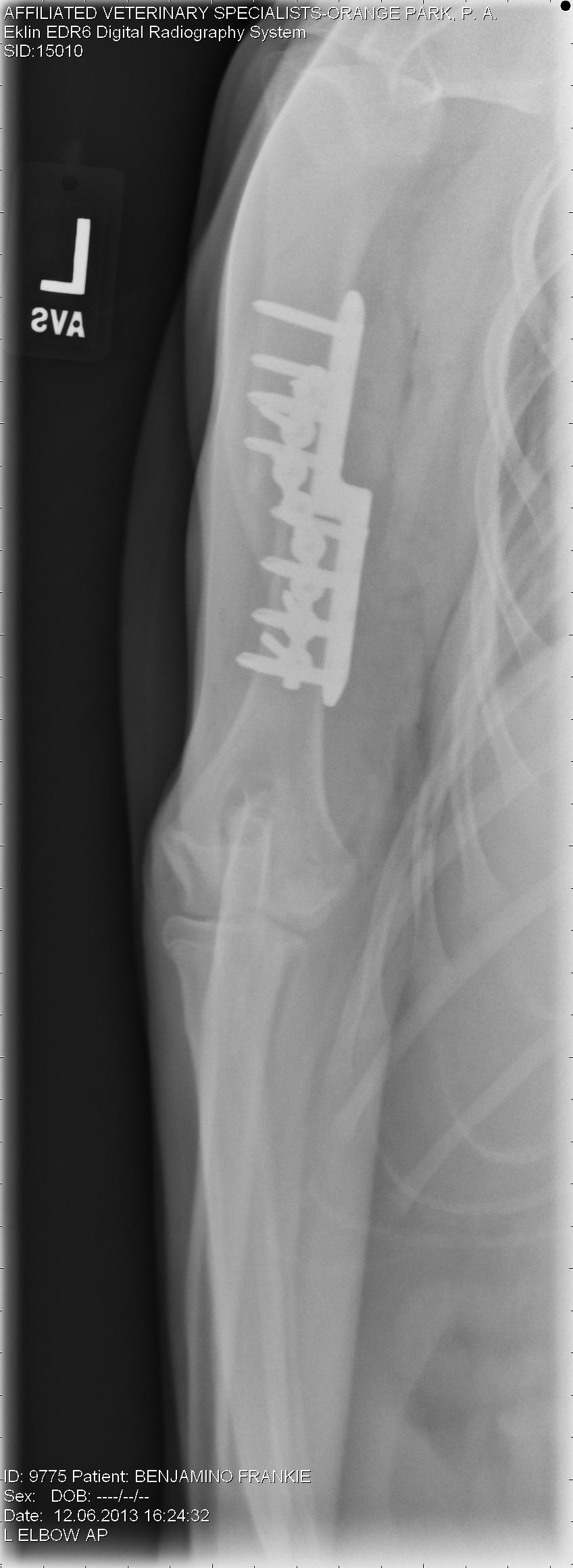
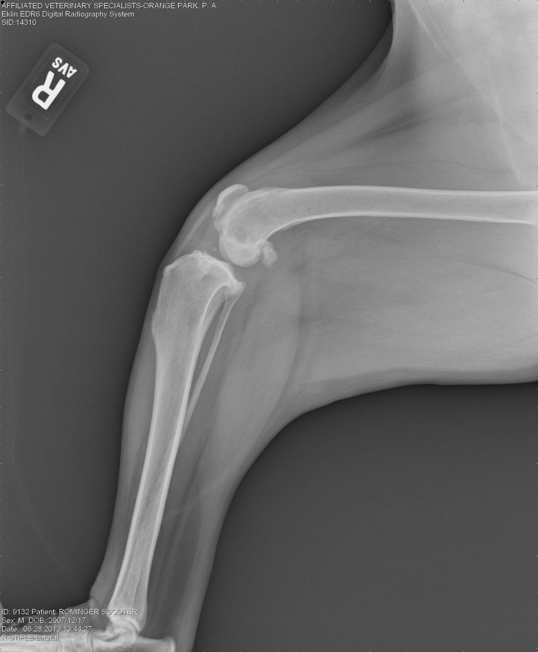
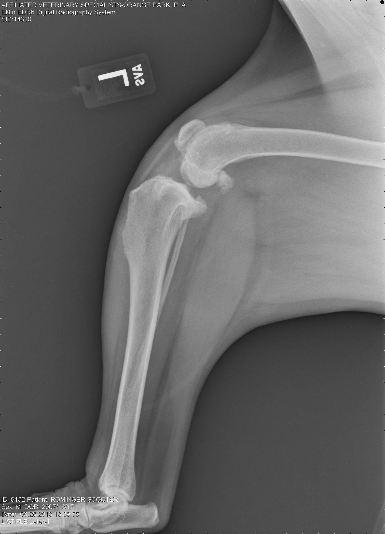
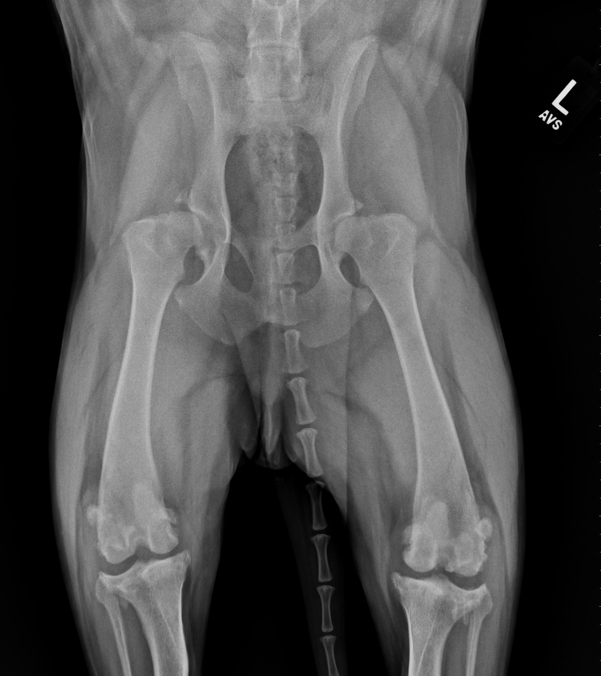
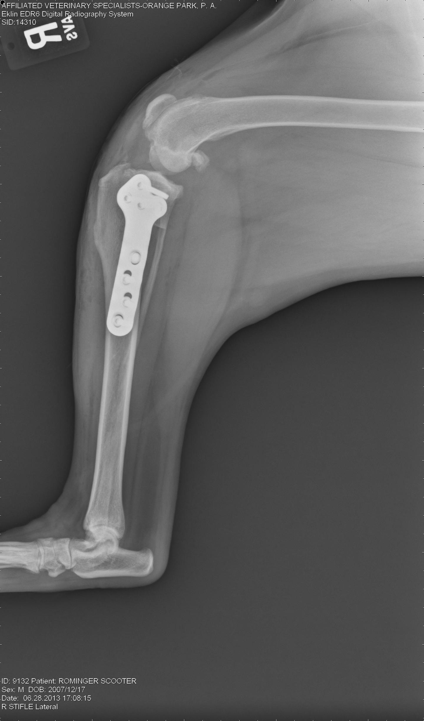
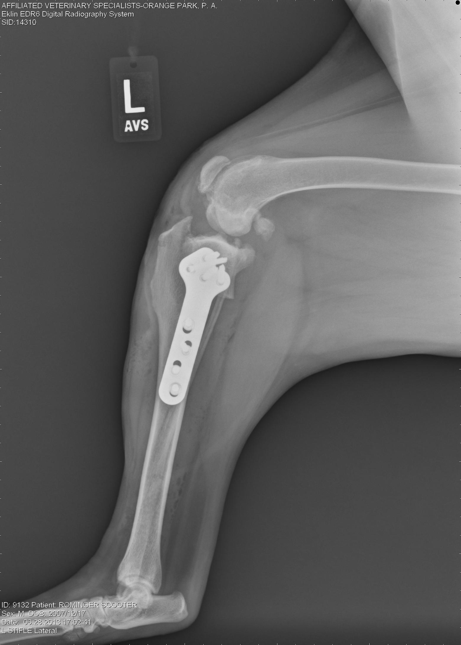
Radiographs (X-rays)

Shoulder_OCD0001 Shoulder_OCD0002

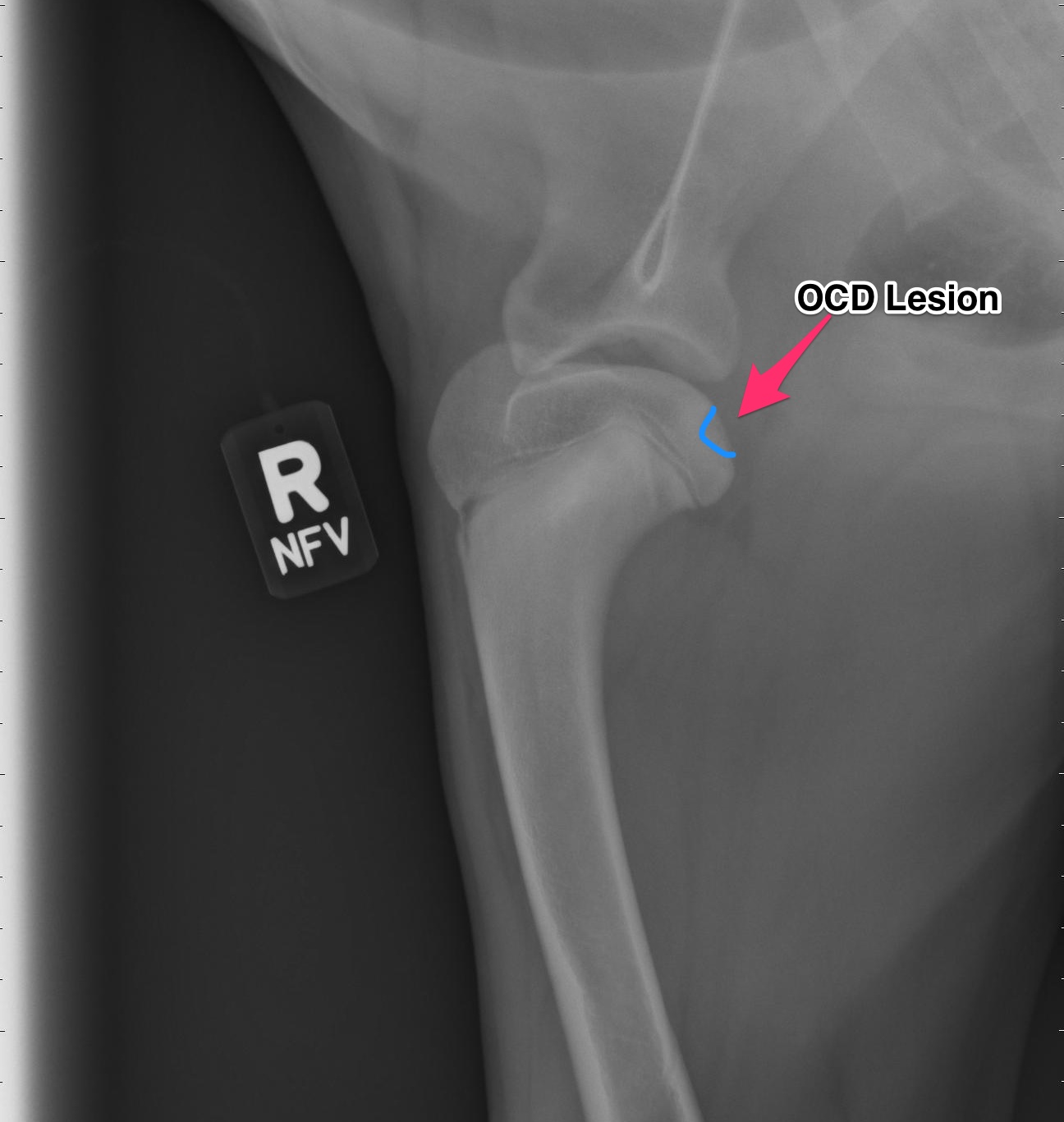
Above are x-rays of a left and right shoulder affected with OCD lesions. These are on the same patient. The images labeled with the left (L) marker has a flattened region noted by the arrow which is characteristic of OCD. The image on the right has the area highlighted in blue. While the lesion doesn’t look big, it can definitely cause a lot of pain and discomfort.

Another way to diagnostically evaluate the joint is with a computed tomography (CT) scan. This will give more detail into the region of interest. Generally this is not needed, however indications for it may be to evaluate the elbows as well.

Treatment:

For the best possible outcome do not delay treatment! At this time, the gold standard approach is arthroscopic debridement (removal) of the fragmented cartilage and the surrounding diseased cartilage and subchondral bone. Curettage may allow the now vacant cartilage bed to fill in more quickly with what is called fibrocartilage. I likened the removal of the fragment to old wallpaper removal (very much oversimplified). Once the old wallpaper bubbles and tears, you need to remove all the damaged wallpaper in the periphery or else the wallpaper will continue to peel.

If the cartilage is an osteochondrosis (OC) lesion and has not fragmented (OCD) non-surgical treatments (activity restriction, dietary restriction, etc) may be attempted and successful. Unfortunately, if OCD has not occurred then the patient will not be limping and most of these dogs go undiagnosed. It is my belief that any dog exhibiting pain/lameness with the presence of a radiographic (x-ray) OCD lesion ,should have surgery. Surgery will benefit them both in the short term and the long term.

There are older techniques of opening the joint to get access to the cartilage flap, however the recovery time on this type of procedure is significantly longer than with arthroscopy. Also, potential complications are increased with an “open” technique than with arthroscopic techniques. Arthroscopy is a minimally invasive tool that allows us to both diagnose and treat this condition. Generally speaking the patient can walk on the surgery leg (even if both legs have surgery at the same time!) following an arthroscopic procedure. Generally 2-3 small ports are placed over the shoulder (2-4mm in length) and this allows us access to the joint and work within the joint.

Recovery and Rehabilitation: 

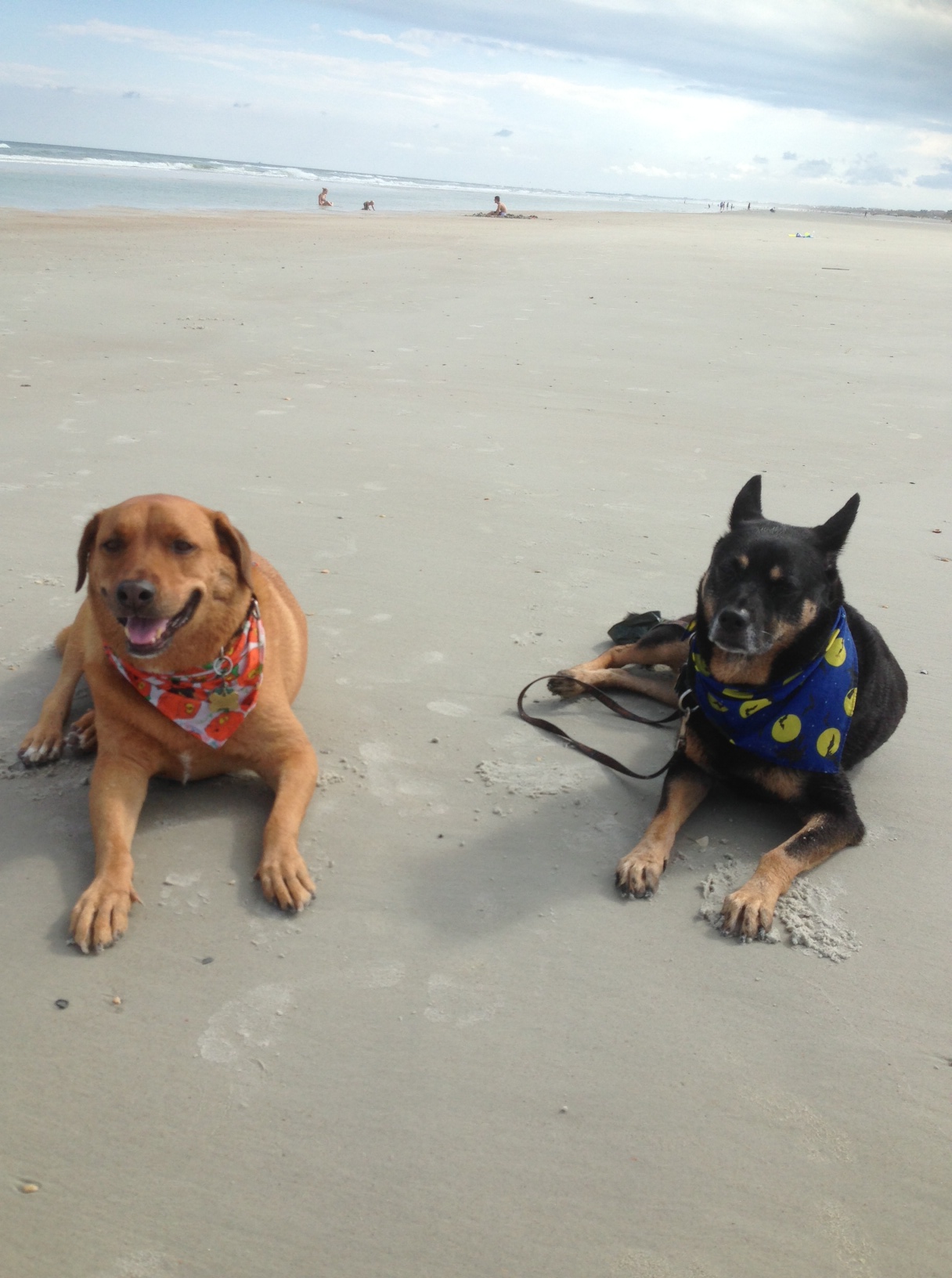
Recovery for the arthroscopic procedure is generally 4-6 weeks. Every surgeon has a different protocol for after surgery and I am very respectful of that. I prefer controlled movement for my patients. In the first two weeks, passive range of motion is very important, followed by active icing of the joint(s). Short leash based walks are started shortly after surgery and incrementally increased as we proceed through the recovery phase. Introduction into a formal rehabilitation program is recommended, however there are times when this is not possible and rehabilitation must be performed at home. Below is a patient that had a single shoulder arthroscopy, you can see how well they can walk following surgery (this is the following day)!

Prognosis:

When diagnosed and treated early, the dog affected with OCD can have a good prognosis and resume a normal or near normal activity level and quality of life. The longer the lesion is present, the more inflammation and arthritis will develop decreasing our success with surgery. Of the OCD lesions (shoulder, versus the other sites affected) this region has the best prognosis. I do encourage all my patients to continue on joint supplementation for life and to be removed from any breeding program.


Frankie’s Journey: Part 2

Frankie relaxing before surgery.

After the diagnostics were performed (reviewed in the previous segment), a decision needed to be made as to which issue was to be addressed first. This is often a tough decision and must be made with the experience of your trusted veterinarian. My criteria consisted of which leg was most affected and the fact that I had already decided to address his hips with a total hip replacement. In my opinion (and supported clinically), a total hip replacement was a better option than other procedures available for canine hip dysplasia. Certain growth plates (centers of bone growth) need to be closed prior to performing a total hip replacement making the minimum age being around 10 months of age. This made our decision easier and the decision to address the elbows was made.

Surgery for his elbows was staged; the left elbow surgery performed on December 6, 2013 and the right elbow addressed on February 14, 2014 (Happy Valentine’s Day!). The time period in between was used for physical therapy and maximizing the results of the surgeries. Identical surgeries were performed on both elbows due to the similarity of the disease process occurring. An elbow arthroscopy was performed, which revealed the cartilage flap (osteochondritis dissecans (OCD)) and a fragmented coronoid process (FCP) The OCD flap was removed and the FCP segment was removed. The damaged bone under the OCD flap was removed with a shaver until healthy bone was present. Since the diseased area of the elbow was on the inside (medial) aspect a specialized procedure to displace weight to the central and outside (lateral) aspects was performed. This procedure is called a sliding humeral osteotomy (SHO). This procedure is fairly aggressive and involves a bone cut in the middle of the humerus bone and a plate/screws placed.

Below are images of the elbow arthroscopy showing both the OCD lesion and the FCP segment.


Noted the line in the cartilage outline a fragmented coronoid Process (FCP).

Noted the line in the cartilage outline a fragmented coronoid Process (FCP).

Note the underlying bone (pink). This is usually covered by cartilage, however the cartilage has become a flap.

Note the underlying bone (pink). This is usually covered by cartilage, however the cartilage has become a flap.


Following the elbow arthroscopy, the SHO procedure was performed. Below is a radiograph of the procedure.

Note the cut in the middle of the humerus and the shifting of the bone.

Note the cut in the middle of the humerus and the shifting of the bone.

Frankie’s Journey

Image

I want to share this story of Frankie with everyone, as it will be an ongoing story for the next year or so as we continue to help him though this hard time. Frankie was seen by me about two months ago when he was six months old. He is a Golden Retriever that presented for lameness in all legs. He had been enrolled in a service dog program when his owners started to realize that he was having trouble walking. He was referred to me after being evaluated by a local neurologist, who couldn’t diagnose a neurologic issue.

On presentation, Frankie had lameness (limping) in all four limbs. He had pain on manipulation of both elbows, especially when pressure was placed on the inside of the elbows. He had a shortened stride to both hind limbs and was painful on hyperflexion and hyperextension of both hips. Another interesting finding, was that both hips could be felt subluxating on exam (positive ortolani test). This means that you could feel the femoral head rub and partially come out of joint.

Radiographs (x-rays) were taken of all joints and a diagnosis of bilateral elbow dysplasia (osteochondrosis dissecans (OCD) and fragmented coronoid process) and bilateral hip dysplasia was made. Unfortunately, Frankie’s career as a service dogs had to abruptly end. Because of the extensive orthopedic work that would be needed (both elbows and both hips) he was in need of a new home to care for his special needs. My wife and I may be a glutton for punishment, but we thought long and hard about this decision and decided to open our home and give this Golden puppy a second chance.

So my intention for this “Featured Article” segment is to follow the course of Frankie’s treatment including surgery, recovery, physical therapy, and final outcome. I feel that other owners may be in similar circumstances and this may help encourage some and educate others. This will also give a forum to discuss congenital issues such as elbow dysplasia and hip dysplasia. Going through these issues on our own pet, has been an eye-opening experience for us and me professionally. I can now relate to my patients and clients on a much more personal level.

In future segments (soon to follow) we will go through diagnosis, diagnostic test (radiographs and CT scan) and surgeries. So far, Frankie has had surgery on one elbow and is recovering well from that, we will go into more specifics as we go. The plan will be for the other elbow in the near future and then total hip replacements.

Also, please do not ask to donate financially to Frankie, I am not trying to raise money. I am just trying to educate others. There are so many generous people out there and there are many charitable animal organizations that can benefit from your generosity, as it is always appreciated!

October 2013 Case of the Month

October Case of the Month

Scooter (left) enjoying the beach!!

Scooter (left) enjoying the beach!!

For October’s Case of the Month, I have selected a relatively common problem that we see in veterinary medicine – cranial cruciate ligament ruptures. Cranial cruciate ligament (CrCL) ruptures (more commonly referred to as an ACL tear after the human literature) are commonly seen in the practice of veterinary surgery, in fact they are our most common orthopedic case that we see. This disorder affects both the large and small dog, from the Great Dane to the Chihuahua and can affect dogs of any age most commonly the middle age dog. If you would like further details about this specific disorder, please see the previous posts regarding cranial cruciate ligament ruptures (click on the orthopedics tab in the menu bar).

Scooter is a  5  yr old Labrador Retriever that presented for lameness in both hind limbs. His history was such that he was lame in the left hind limb about a year ago and had a previous surgical procedure to address the CrCL performed, to which he responded well early on but became increasingly lame again in the leg and then developed a right hind limb lameness in addition. The procedure previously performed on the left stifle (knee) was not documented and no radiographic implants were used in or around the stifle. Also, Scooter has a chronic history of hip dysplasia and osteoarthritis in both hips to compound his issues.

Physical Exam:

Scooter could walk with assistance, however really struggled in both hind limbs to ambulate. Also, you could see Scooter shifting his weight to his front legs, which is a very classic feature for dogs with CrCL ruptures that affects both stifles. Our physical exam revealed that both (left and right) CrCL were ruptured and we highly suspected bilateral meniscal injuries/tears. While some discomfort could be elicited from manipulation of his hips, the majority of his discomfort and inability to walk was from his CrCL ruptures and meniscal tears.

Right knee - note the joint swelling, arthritic changes, and forward movement of the tibia in relation to the femur.

Right knee – note the joint swelling, arthritic changes, and forward movement of the tibia in relation to the femur.

Left knee - note the joint swelling, arthritic changes, and forward movement of the tibia in relation to the femur.

Left knee – note the joint swelling, arthritic changes, and forward movement of the tibia in relation to the femur.

VD pelvis x-ray - note the chronic signs associated with hip dysplasia.

VD pelvis x-ray – note the chronic signs associated with hip dysplasia.

Surgery:

Surgery was scheduled soon after his initial exam, all his pre-operative work-up was otherwise normal. Most of the time we try to stage each leg. The big reason for separating out surgery on each leg is to reduce the risk of complications such as infection and implant breakdown. Some cases, like Scooter, we chose to do both especially if they are severely affected on both legs like Scooter.

At surgery, bilateral cranial cruciate ligament ruptures were noted, along with bilateral medial meniscal tears. All those findings can be very painful for the patient. Both meniscal tears were debrided (removed) and bilateral tibial plateau leveling osteotomies (TPLOs) were performed. For more detailed information about ways we correct CrCL tears, please view that page on this website.

Right knee - following TPLO surgery.

Right knee – following TPLO surgery.

Left knee - Following TPLO surgery.

Left knee – Following TPLO surgery.

Post-operative care:

As you can image, we treat these patients very carefully. In human medicine, physical therapy and rehabilitation is started almost immediately following surgery. As soon as a patient leaves the operating room, we start icing of the surgical site. That is followed but passive range of motion exercises and short, assisted walks and frequent icing after sessions during the first two weeks. A fairly strict physical therapy program is given to owners and in some cases, organized physical therapy sessions are scheduled under the supervision of a certified canine rehabilitation therapist (CCRT). I generally tell the owners that their commitment to physical therapy is as important as the surgery performed. In Scooter’s case, his owners were very dedicated to the whole process and 16 weeks later he is back to doing his normal activity, which includes running, swimming, and of course lounging around from time to time.

Swimming at dusk.

Swimming at dusk.

Happy dog basking in the sun!!

Happy dog basking in the sun!!

Scooter and his buddy enjoying a swim!!

Scooter and his buddy enjoying a swim!!

Happy Holidays

I want to take a moment to thank everyone for their continued interest and support of this blog. Your continued support has meant a lot to me and allowed me to continue to post various topics. I want to wish everyone Happy Holidays during this joyous time of the year! I wish you all the best in the New Year too! Keep your pets safe throughout this time, but definitely spoil them (we want them to ring in the New Years without any ailments)! Please continue to follow this blog!

Seasons Greetings!!

Kevin

Minimally Invasive Surgery – Laparoscopy

View of the liver and diaphragm laparoscopically

View of the right ovary during a standard laparoscopic spay.

Abdominal view of a laparoscopic gastropexy.

What is laparoscopy? This is an advanced surgical technique that utilizes cameras and instruments that are introduced into the abdomen via small incisions (portals). This type of surgical procedure is becoming more and more common in human medicine. It is not uncommon to hear about gallbladder removals, “lap band” procedures and many other operations being performed this way. It is really becoming the standard of care – so why not for our pets? Certainly the technology is there! The more and more we get comfortable with these techniques, the more we will be able to do.

The basic concept of laparoscopy is to make portals that allow the introduction of a camera and instruments. The actual surgical work is done within the abdomen by visualizing it on a video monitor. Before attempting laparoscopic surgeries, the surgeon needs to be comfortable with performing the surgery in an open approach, because there are times, when laparoscopy is not the best approach and the surgery needs to be converted.

What are the benefits of laparoscopy? The most notable benefits are patient comfort, soft tissue damage (termed morbidity), excellent visualization (images are magnified), shorter hospital stays. All these combined form a convincing argument for laparoscopic surgery.

One of the most common procedures performed is the ovariohysterectomy (spay). This can be performed from 0.5 cm and 1.0 cm incisions. This is a very quick procedure and allows the surgeon complete access and visualization of the ovaries and uterus. Specialized sealing devices allow the surgeon to remove the full uterus and ovaries with minimal bleeding and incisions. Another benefit is that with the small size of the incisions, there is low risk of herniation of abdominal contents, and the patient can return to activity in about a week.

The spay is many times combined with a prophylactic gastropexy in the larger breed dogs. This is a procedure that allows the stomach to be attached to the body wall to prevent deadly twisting of the stomach. This is a very effective procedure. For more information, please visit the previous blogs entitled GDV.

Other more complex procedures include multiple biopsies, adrenalectomy, gallbladder removal, liver mass resection, cryptorchid testicle removal, etc. As more experience is gained with laparoscopy the more we can do. If you have questions about laparoscopy feel free to comment or ask your veterinarian and veterinary surgeon.

Minimally Invasive Surgery – 1

I hope everyone is having a great weekend! The next series of topics is join got focus on minimally invasive procedures. This is of particular interest of mine. There are multiple implications of this terms, I am going to focus on laparascopy, thoracoscopy and arthroscopy.

As medicine advances in both the human and veterinary world, we are starting to do more with less. What I mean is that through smaller incisions and better technology we address more and more surgical problems. The benefit of this is shorter hospital stays, decreased complication rte, decreased infection rates, and decreased soft tissue trauma and post-surgical pain. We are very lucky to have all this new technology available to us.

Here is a definition of the terms:

Laparascopy: Abdominal surgery being performed with a scope/camera via a small portal incision. Multiple other portals are made to introduce instruments. All procedures are performed within the abdominal cavity. Laparoscopic-assisted means that part of the procedure is done with cameras within the body cavity and part is done via small incisions outside of the abdomen.

Thoracoscopy: Same definition as above, however this pertains to the chest or thorax. This is a very useful modality and is used to treat various cardiac, pulmonary (lung) and esophageal problems.

Arthroscopy: Same definition as above, however this is performed on the joints. This is a very common use in veterinary medicine and is thought to be the gold standard in joint evaluation. The most common joints evaluated through this are the elbows, shoulders and knees, however the wrists (carpi), ankles (hocks/tarsi), and hips can be evaluated. Just think, if you have an ACL injury, it is very common for you to have your knee scoped, why not your best friend?